ORIGINAL RESEARCH

Evaluation of efficacy of the amino acid-peptide complex administered intragastrically to golden hamsters experimentally infected with SARS-CoV-2

Laptev DS1, Protasova GA1, Petunov SG1, Radilov AS1, Chepur SV2, Gogolevskiy AS2, Myasnikov VA2, Tyunin MA2, Smirnova AV2
About authors

1 Research Institute of Hygiene, Occupational Pathology and Human Ecology Leningrad Region, Russia

2 State Research and Testing Military Medicine Institute under the Ministry of Defense of the Russian Federation, St. Petersburg, Russia

Correspondence should be addressed: Denis S. Laptev
stancija Kapitolovo, stroenie 93, r.p. Kuzmolovsky, Vsevolozhsky r., 188663; ur.liam@nedpal

About paper

Author contribution: Laptev DS, Protasova GA, Myasnikov VA, Tyunin MA, Smirnova AV — experiment, information collection, data processing; Petunov SG — data processing and interpretation; Radilov AS — scientific concept, consulting; Chepur SV — experiment organization, COVID-19 in vivo model development; Gogolevskiy AS — experiment organization. All authors participated in the manuscript authoring and editing.

Compliance with ethical standards: the study was conducted in conformity to the provisions of the European Convention for the Protection of Vertebrate Animals used for Experimental and other Scientific Purposes.
Received: 2021-05-21 Accepted: 2021-06-06 Published online: 2021-06-16
|
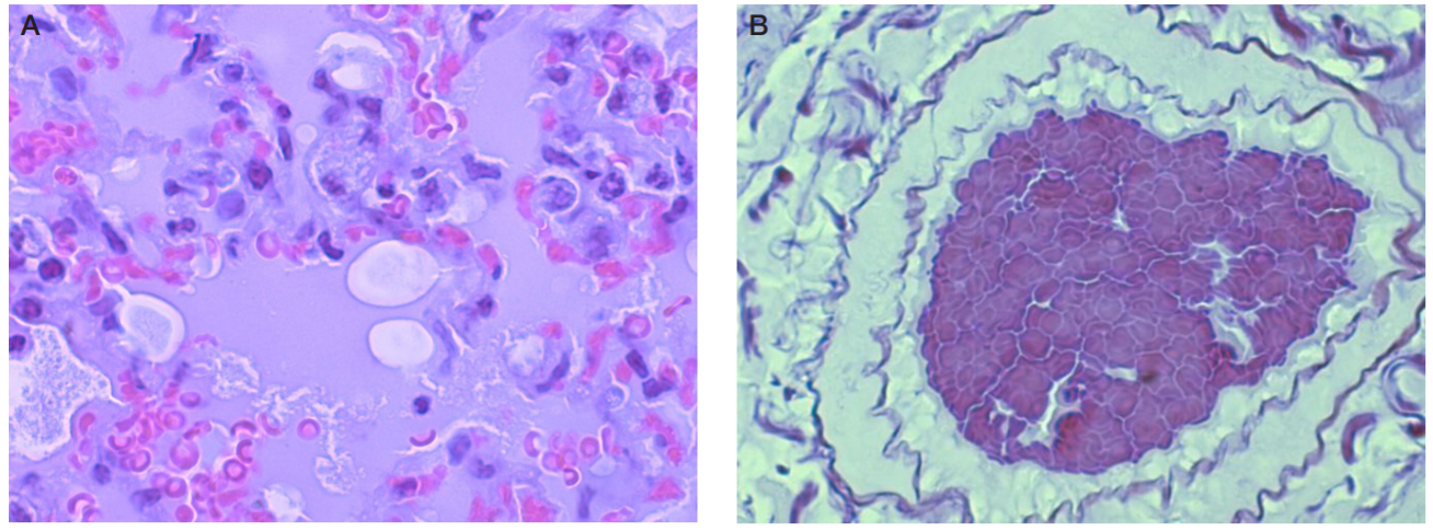
Fig. 1. Lung. Group 2. A. Alveolar edema (stained with hematoxylin and eosin; magnification ×1000). B. Thrombosis (weigert staining for fibrin; magnification ×650)
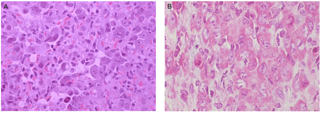
Fig. 2. Lung. Group 2. A. Diffuse alveolar damage, hypertrophied atypical type II alveolocytes (hematoxylin and eosin staining; magnification ×650). B. RNA of the cytoplasm of atypical type II alveolocytes stained pink-red, Brachet staining (pyronine and methyl green; magnification ×1000)
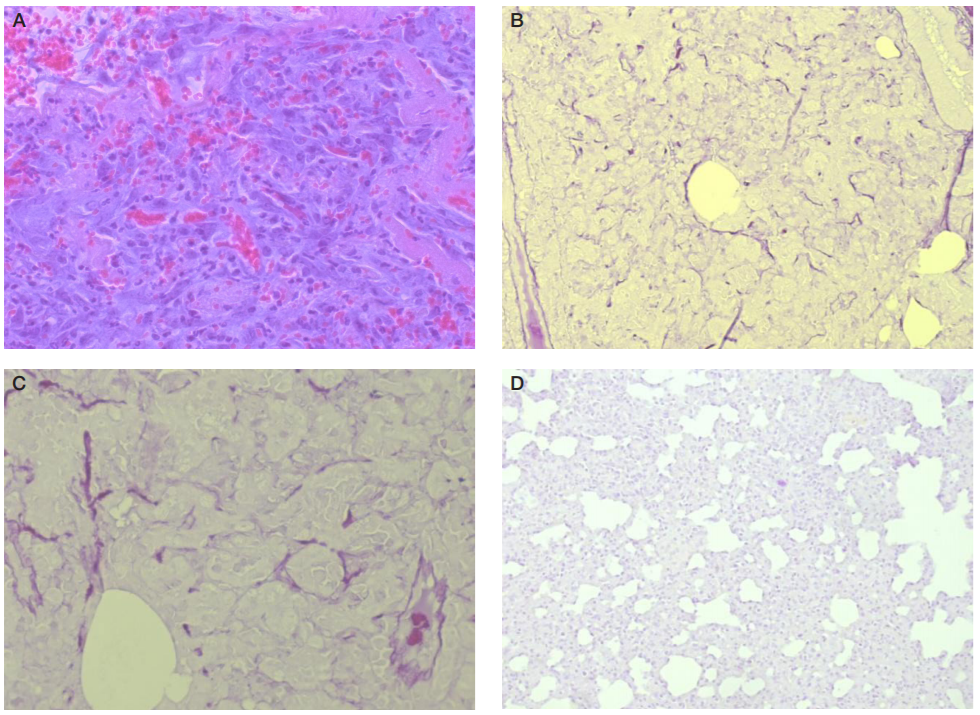
Fig. 3. Lung. Group 2. A. Fibroblast proliferation, focal fibrosis, hemorrhage (staining with hematoxylin and eosin; magnification ×400). B. Diffuse deposition of fibrin in the parenchyma (magnification ×200). C. Fibrin deposits in lung parenchyma (magnification ×400). D. Group 3, no fibrin (weigert staining for fibrin; magnification ×200)
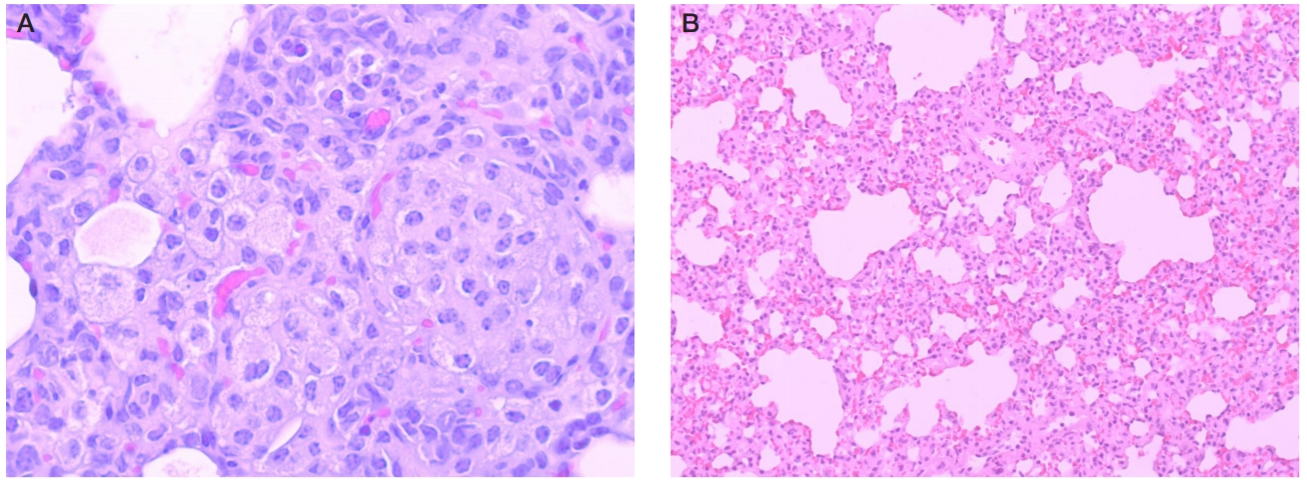
Fig. 4. Lung. Group 3. A. Polymorphic-cell infiltration, reinforced macrophageal response (magnification ×650). B. No diffuse alveolar damage in the lungs (staining with hematoxylin and eosin; magnification ×200)
Table 1. Body weight of Syrian hamsters infected with SARS-CoV-2; absolute and relative data presented as М ± SE (n = 10)
Note: # — statistically significant difference from the background; * — statistically significant difference from group 1.
Table 2. Mass ratios of internal organs of Syrian hamsters infected with SARS-CoV-2 (М ± SE; n = 10)
Note: * — statistically significant difference from group 1; # — statistically significant difference from group 2.