ORIGINAL RESEARCH

Computational phantom for red bone marrow dosimetry from incorporated beta emitters in a newborn baby

About authors

1 Urals Research Center for Radiation Medicine of the Federal Medical-Biological Agency, Chelyabinsk, Russia

2 Chelyabinsk State University, Chelyabinsk, Russia

Correspondence should be addressed: Pavel A. Sharagin
Vorovskogo, 68-а, Chelyabinsk, 454141, Russia; ur.mrcru@nigarahs

About paper

Funding: The work was performed within the framework of the Federal Targeted Program "Nuclear and Radiation Safety" and was financially supported by the Federal Medical — Biological Agency of Russia. The methodological approaches were developed with financial support from the Federal Medical — Biological Agency of Russia and the Office of International Health Programs of the U.S. Department of Energy as part of the joint U.S.-Russian JCCRER 1.1 project.

Author contribution: all authors contributed equally to the development of research methodology, data acquisition, analysis, and interpretation, and to the writing and editing of the article.

Received: 2022-11-10 Accepted: 2022-12-19 Published online: 2022-12-27
|
Fig. 1. Newborn’s iliac phantom section (trabeculae’s and cortical bone are shown in black , red bone marrow is shown in white )
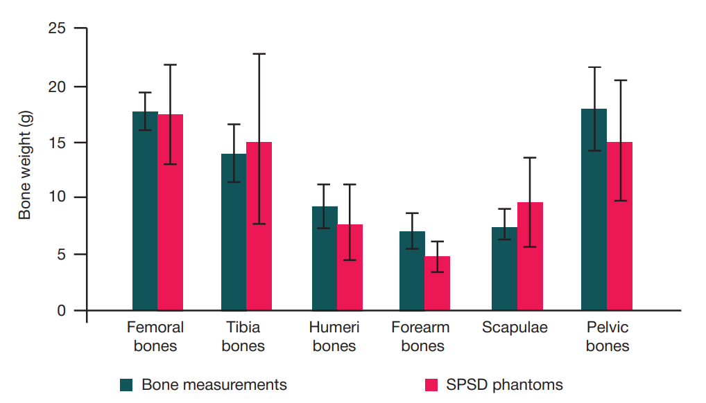
Fig. 2. Comparison of measured bone masses [68], and masses corresponding to the sizes of SPSD phantoms estimated as the sum of masses of all segments describing specific bones (left + right) of newborns, taking into account repeating and paired segments. Error bars showed the standard deviations."
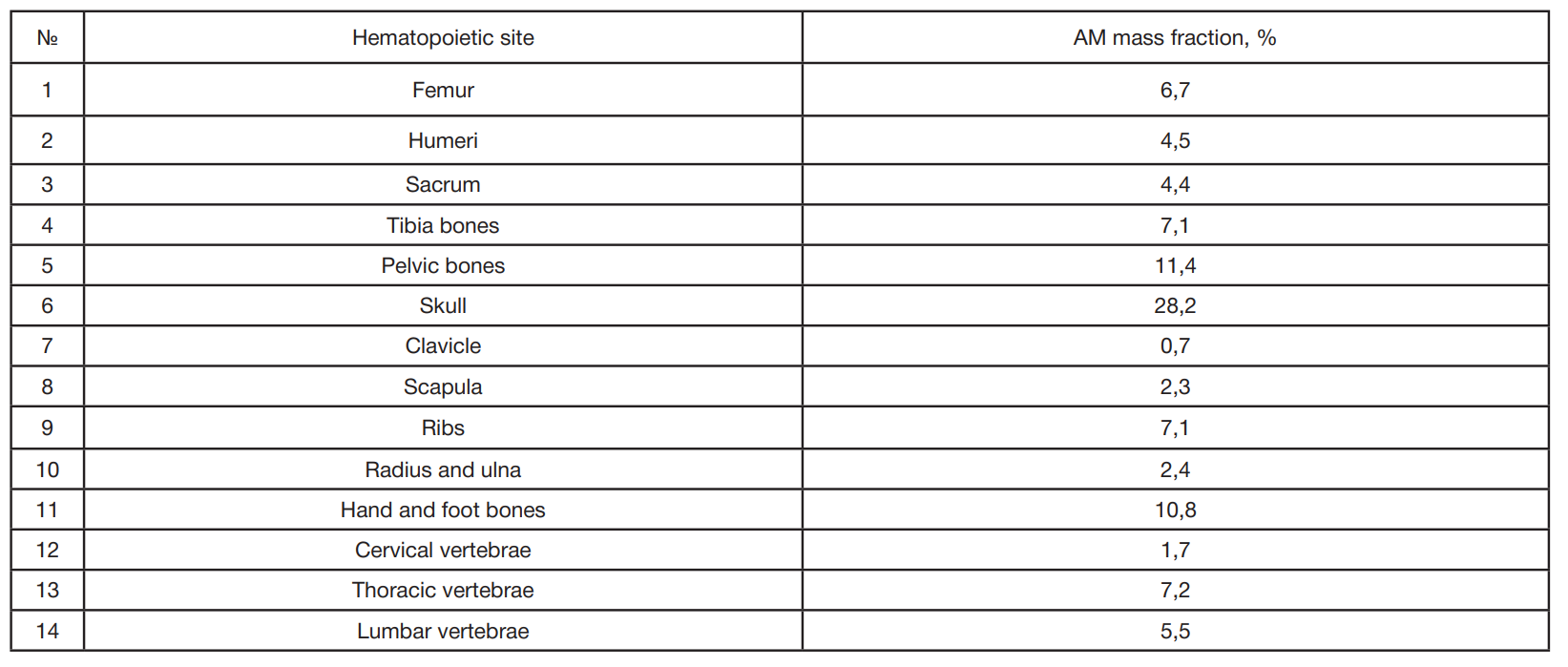
Table 1. Mass fraction of AM (% of the total mass of AM in the skeleton) in the main hematopoietic sites of the skeleton of a newborn baby [17]
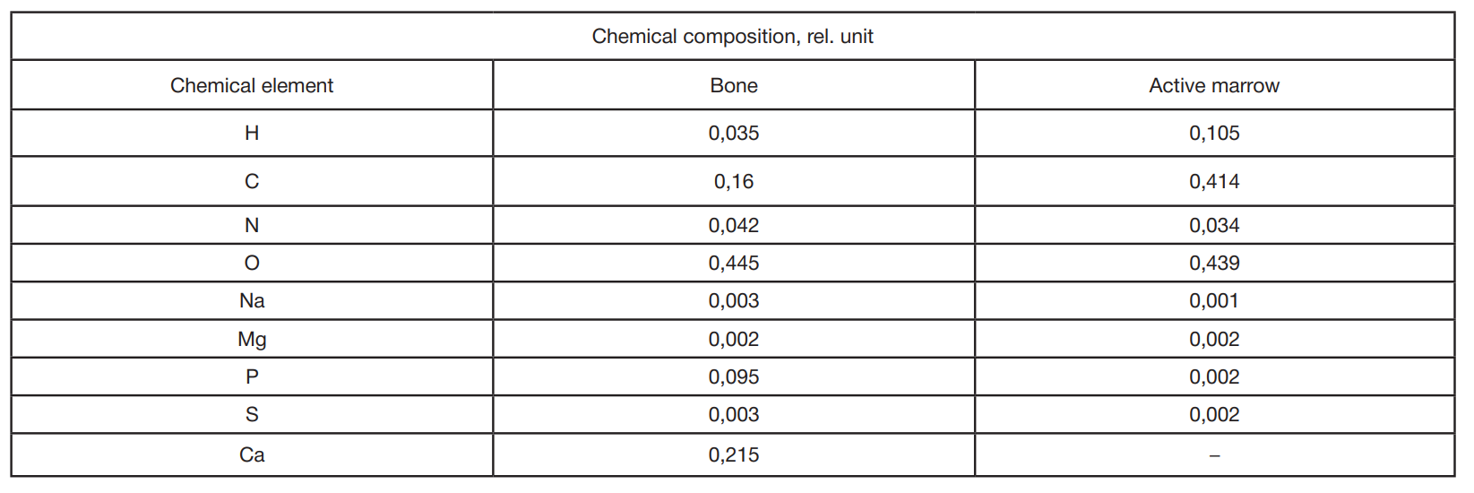
Table 2. Chemical composition of simulated media adopted for all BPS
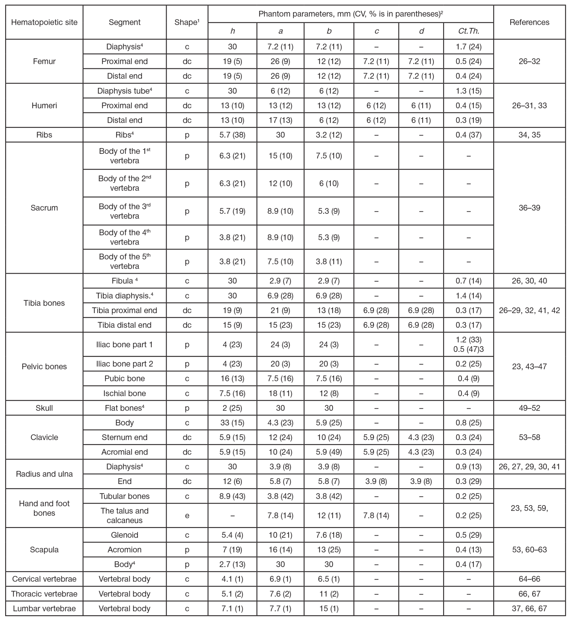
Table 3. Linear dimensions and cortical thickness accepted for BPS of a newborn baby
Note: 1 — the shape of the phantom was designated as follows: c — cylinder, dc — deformed cylinder, p-rectangular parallelepiped, e — ellipsoid; 2 — the dimensions of the BPS were designated as follows: h — height; a — major axis (c), major axis for a larger base (dc), or side a (p); b — minor axis (c), minor axis for a larger base (dc), or side b (n); c — major axis for a smaller base (dc); d — minor axis for a smaller base (dc); for an ellipsoid (e), a, b, c denote the axes of the ellipsoid; 3 — the thickness of the cortical layer was taken to be different for the inner (medial) and outer (gluteal) surfaces of this segment of the iliac bone (Fig. 1); 4 — the BPS imitated only a part of the simulated bone segment, if the dimensions of the bone segment significantly exceeded 30 mm, since in such cases, from the point of view of dosimetry, it does not have it makes no sense to model the entire bone section as a whole [11, 12].