ISSN Print 2713-2757
ISSN Online 2713-2765
SCIENTIFIC AND PRACTICAL REVIEWED JOURNAL OF FMBA OF RUSSIA
1 Federal Clinical Center for High Medical Technologies of the Federal Medical Biological Agency, Moscow, Russia
2 National Medical Research Center for Otorlaryngology of Federal Medical Biological Agency, Moscow, Russia
3 Academy of Postgraduate Education, Federal Scientific and Clinical Centre for Specialized Types of Medical Care and Medical Technologies of the Federal Medical Biological Agency, Moscow, Russia
Correspondence should be addressed: Arbak А. Khachatryan
Volokolamskoe shosse, 30, bld. 2, Moscow, 123182, Russia; ur.xednay@kabrard
Author contribution: Danishchuk OI, Nazarian DN — surgical procedure, manuscript writing and editing; Karpova EI — surgical procedure; Khachatryan AA — manuscript writing; Razmadze SS — patient management, manuscript writing.
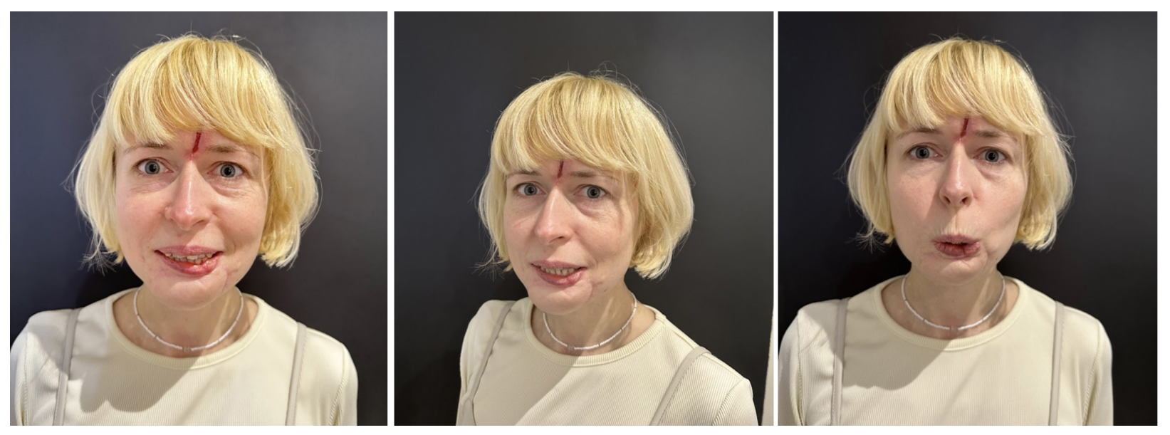
Compliance with ethical standards: the informed consent to case study publication was submitted by the patient.