ORIGINAL RESEARCH

Changes in some immunological parameters after COVID-19: general trends and individual characteristics

Glazanova TV, Shilova ER, Efremova YS, Chubukina JB, Bessmeltsev SS
About authors

Russian Hematology and Transfusiology Research Institute of the Federal Medical Biological Agency, St. Petersburg, Russia

Correspondence should be addressed: Tatyana Valentinovna Glazanova
2-ya Sovetskaya, 16, St. Petersburg, 191023, Russia; ur.xednay@avonazalg-anaytat

About paper

Compliance with the ethical standards: the study was approved by the Ethics Committee of the Russian Hematology and Transfusiology Research Institute of the FMBA of Russia (Minutes #31 of July 20, 2023). All participants have voluntarily signed informed consent forms.
Received: 2024-04-02 Accepted: 2024-06-08 Published online: 2024-06-30
|
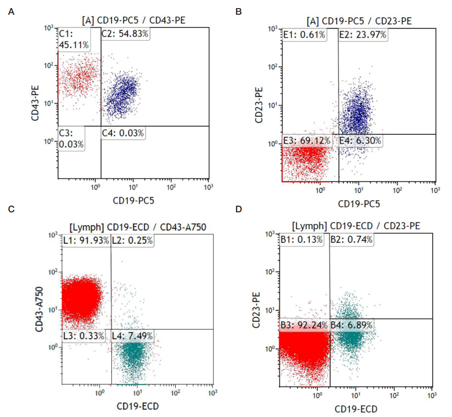
Fig. Immunophenotypic study results, patient A. Device used for the study: Navios EX 10 Colors (Beckman Coulter; USA). The lymphocytic pool was isolated by the parameters of direct and lateral light scattering with CD45 gating. A, B. Results of the study of August 2021. For these histograms, a 5 color panel was used: 1) CD20–FITC+CD23–PE+CD45–ECD+CD19–Pc5+CD5–Pc7 and 2) CD22–FITC+CD43-PE+CD19–Pc5+CD45–Pc7. C, D. Results of the study of April 2023. For these histograms, a 10 color panel was used: FMC7–FITC+CD23–PE+CD19–ECD+CD79b–Pc5.5+CD200–Pc7+CD43–A750+CD38–A700+IgM–PB+CD45–KO. X-axis: CD19+ (%), Y-axis: A and C — CD43+ (%), B and D — CD23+ (%). The upper right quadrants of all the histograms (C2 (A), E2 (B), L2(C) and B2 (D)) show the region where the clone of pathological cells characteristic of B-CLL should be located
Table 1. Post-COVID cellular immunity indicators, groups by disease course severity
Table 2. Post-COVID abnormalities in the CD3+ and CD19+ counts (peripheral blood), % of the examined
Table 3. Content of CIC in COVID-19 patients, depending on the severity of the disease
Table 4. Frequency of occurrence of certain symptoms in COVID-19 survivors depending on the level of CIClow