ORIGINAL RESEARCH

An experiment on biological objects: composite facial graft cross-transplantation

Daykhes NA1, Nazaryan DN1, Gileva KS2, Mokhirev MA1, Lyashev IN1, Zakharov GK1, Fedosov AV1, Potapov MB1, Batyrev AV1, Karneeva OV1
About authors

1 Federal Scientific and Clinical Center of Otorhinolaryngology under the Federal Medical-Biological Agency (FMBA) of the Russian Federation, Moscow, Russia

2 Petrovsky Russian Scientific Center for Surgery, Moscow, Russia

Correspondence should be addressed: Aleksey V. Batyrev
Volokolamskoe shosse, 30/2, k. 443, Moscow, 123182; ur.xednay@laicafoinarc

About paper

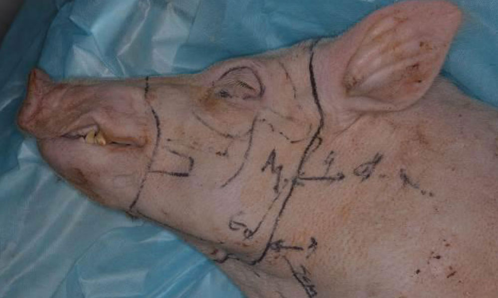
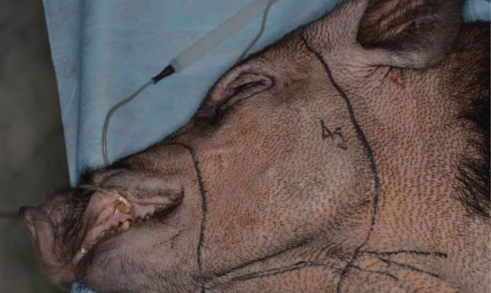
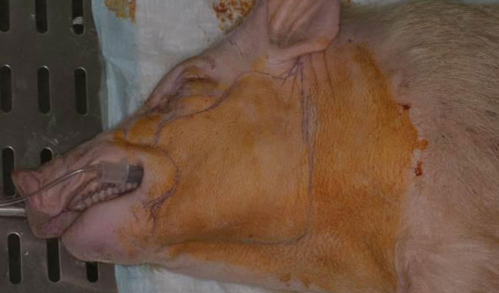
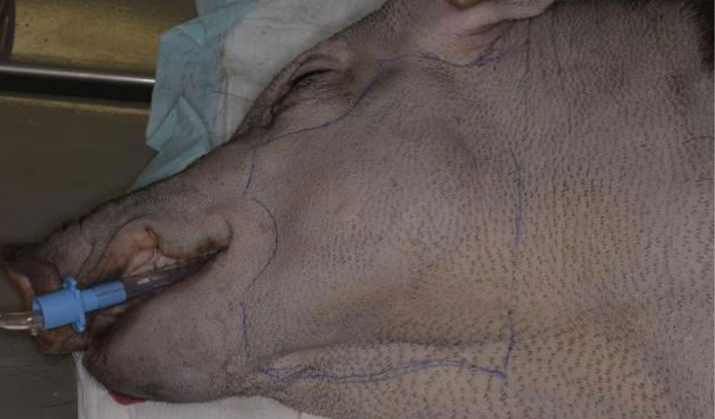
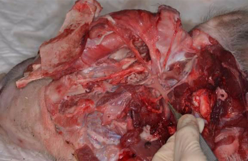
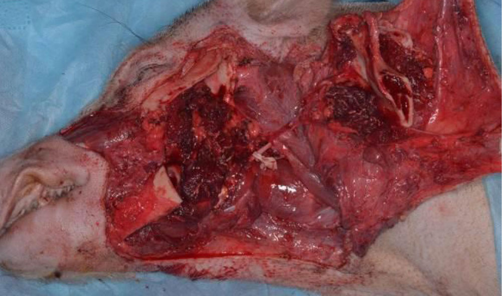
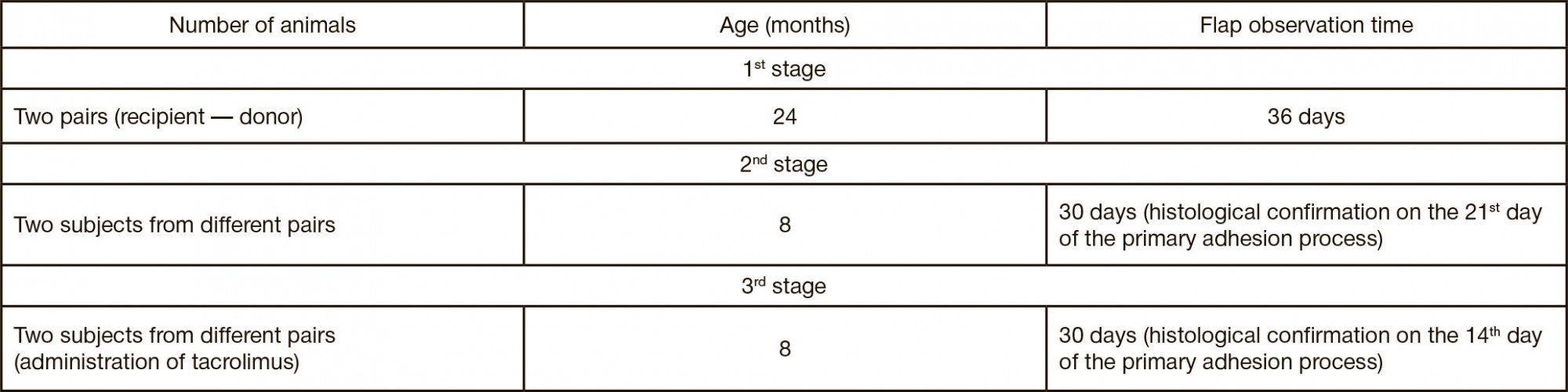
Funding: FMBA applied research, subject "Research of metabolic, morphometric and functional characteristics of tissues and organs after head and neck area surgery involving physical and laser-conversion digital technologies" ("ChLH-18").

Author contribution: Daikhes NA, Nazaryan DN — work organization, article editing; Gileva KS, Mokhirev MA, Lyashev IN, Zakharov GK, Fedosov AV, Potapov MB — participation in the experimental part of the work; Batyrev AV — participation in the organization and experimental part of the work, article authoring; Karneeva OV — participation in the organization of work.

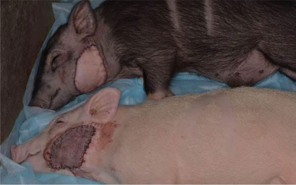
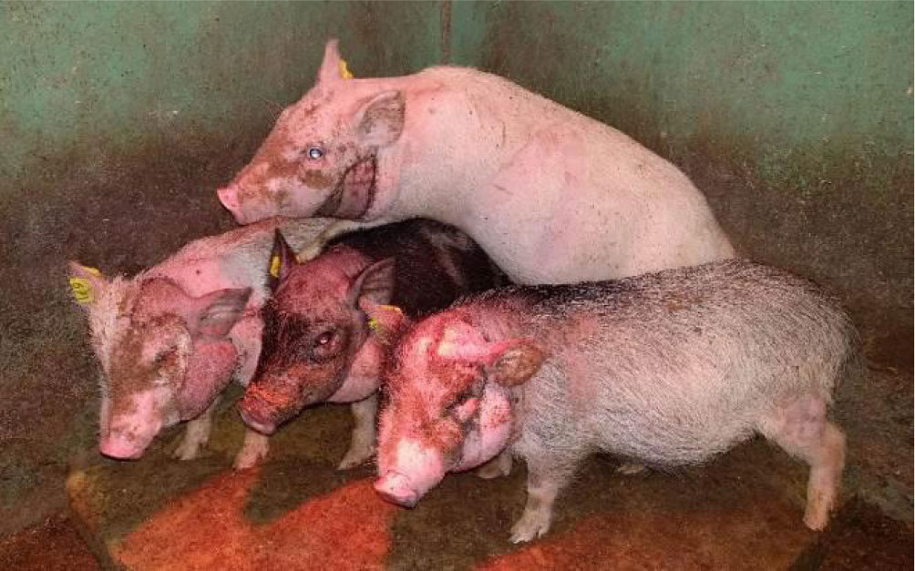
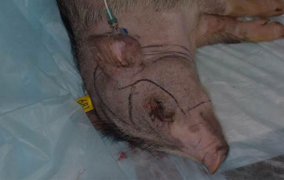
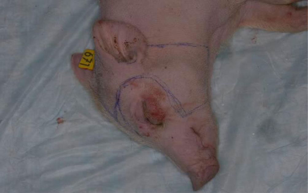
Compliance with ethical standards: the living conditions of animals, care and all manipulations they were subjected to meet the experimental model research standards.

Received: 2020-10-01 Accepted: 2020-11-14 Published online: 2020-11-29
|
Fig. 1. Experimental animals in the immediate postoperative period
Fig. 2. Minipigs on the 14th day after the cross-transplantation
Fig. 3. Facial musculocutaneous flap from the buccal, parotid regions (first subject)
Fig. 4. Facial musculocutaneous flap from the buccal, parotid regions (second subject)
Fig. 5. Composite skin-musculoskeletal flap from the buccal, parotid regions and the lower jaw (first subject)
Fig. 6. Composite skin-musculoskeletal flap from the buccal, parotid regions and the lower jaw (second subject)
Fig. 7. Facial musculocutaneous flap from the buccal, parotid, lower paraorbital regions (first subject)
Fig. 8. Facial musculocutaneous flap from the buccal, parotid, lower paraorbital regions (second subject)
Fig. 9. Facial musculocutaneous flap from the parotid region with auricle and buccal part (first subject)
Fig. 10. Facial musculocutaneous flap from the parotid region with auricle and buccal part (second subject)
Fig. 11. Intraoperative picture taken after dissection of the composite skinmusculoskeletal flap from the buccal region
Fig. 12. Intraoperative picture taken after dissection of the composite skinmusculoskeletal flap from the buccal region and the lower jaw
Fig. 13. Intraoperative picture taken after vascular anastomoses were made
Fig. 14. Intraoperative picture taken after exposition of the facial nerve's trunk and branches
Fig. 15. Place of fusion of the flap with the recipient's tissues on the scanned image of the histological specimen
Table 1. Flap designs used at different stages of the experiment
Table 2. Follow-up time at each stage of the experiment
Table 3. Survival of the animals after surgery
Table 4. Flap survival depending on the type of antibacterial and immunosuppressive therapy selected