ORIGINAL RESEARCH

Using experimental ex vivo models to develop COVID-19 pathogenetic therapy and complications prevention agents

About authors

Research Institute of Hygiene, Occupational Pathology and Human Ecology Leningrad Region, Russia

Correspondence should be addressed: Denis S. Laptev
st. Kapitolovo, gor. pos. Kuzmolovsky, 93, Vsevolozhsky rajon, 188663; ur.liam@nedpal

About paper

Author contribution: Laptev DS — experimental part, information collection, data processing; Petunov SG — data processing and interpretation, general guidance; Nechaykina OV — experimental part, information collection; Bobkov DV — data processing; Radilov AS — data processing and interpretation.

Compliance with ethical standards: all work with animals was carried out in conformity to the provisions of the European Convention for the Protection of Vertebrate Animals used for Experimental and other Scientific Purposes.
Received: 2020-09-15 Accepted: 2020-12-25 Published online: 2020-12-12
|
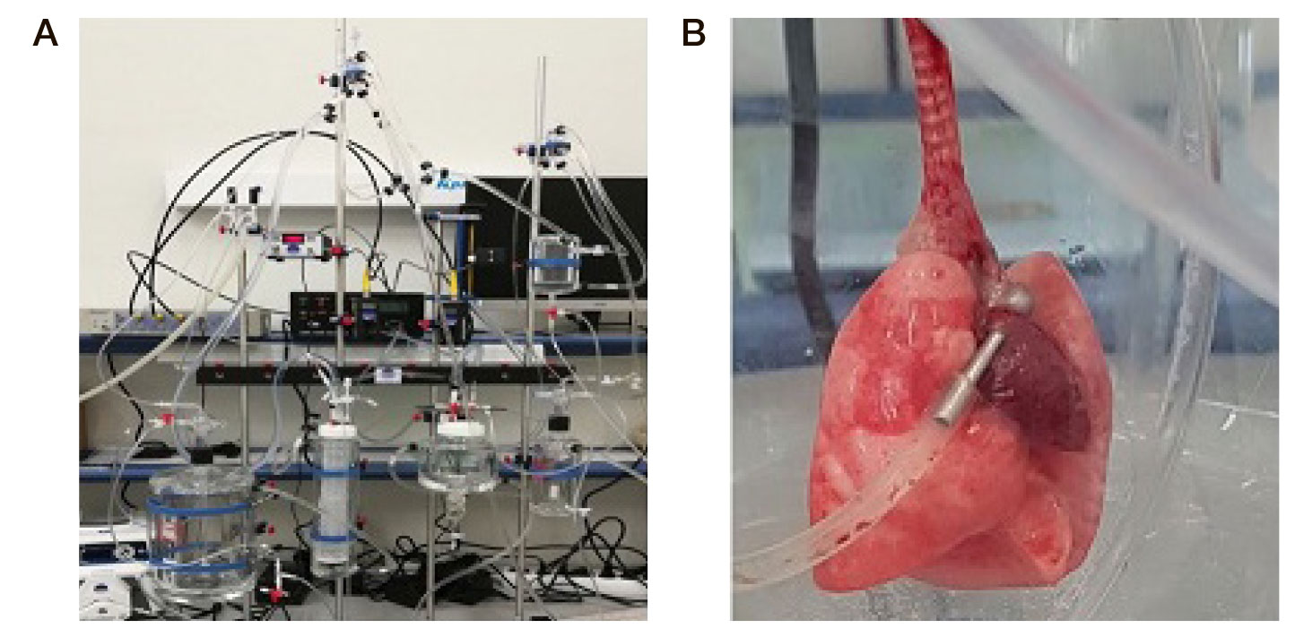
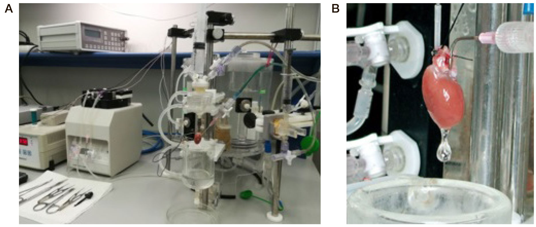
Fig. 1. Isolated lung system. A. General view of the installation. B. Heart-lungs complex
Fig. 2. Installation of an isolated heart according to Langendorff. A. General view. B. Cannulated heart
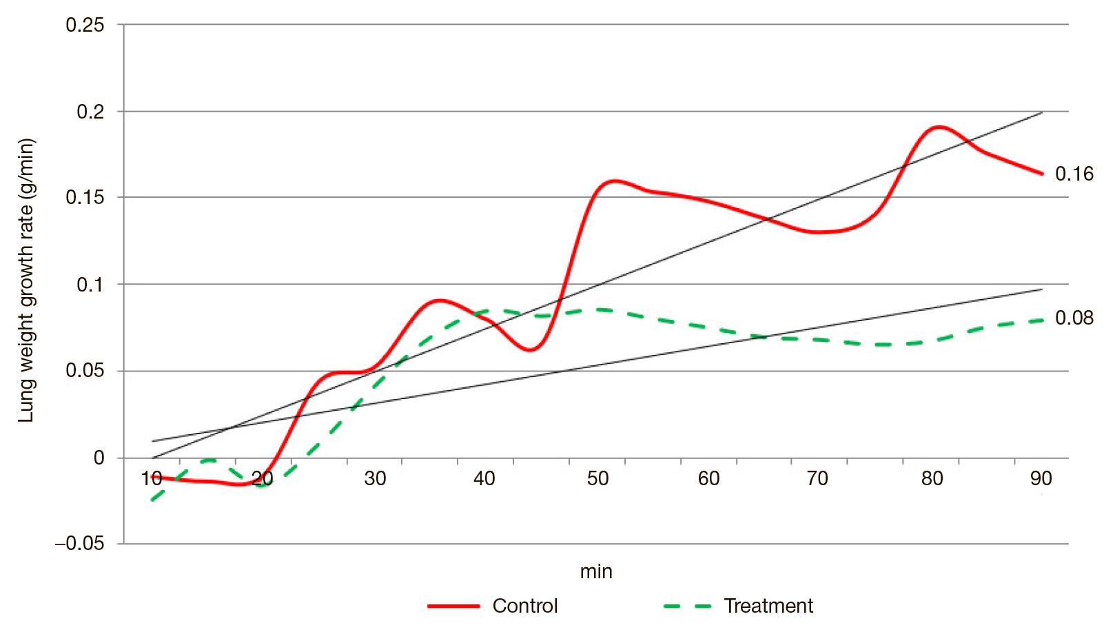
Fig. 3. Lung weight growth rate, treatment and control groups
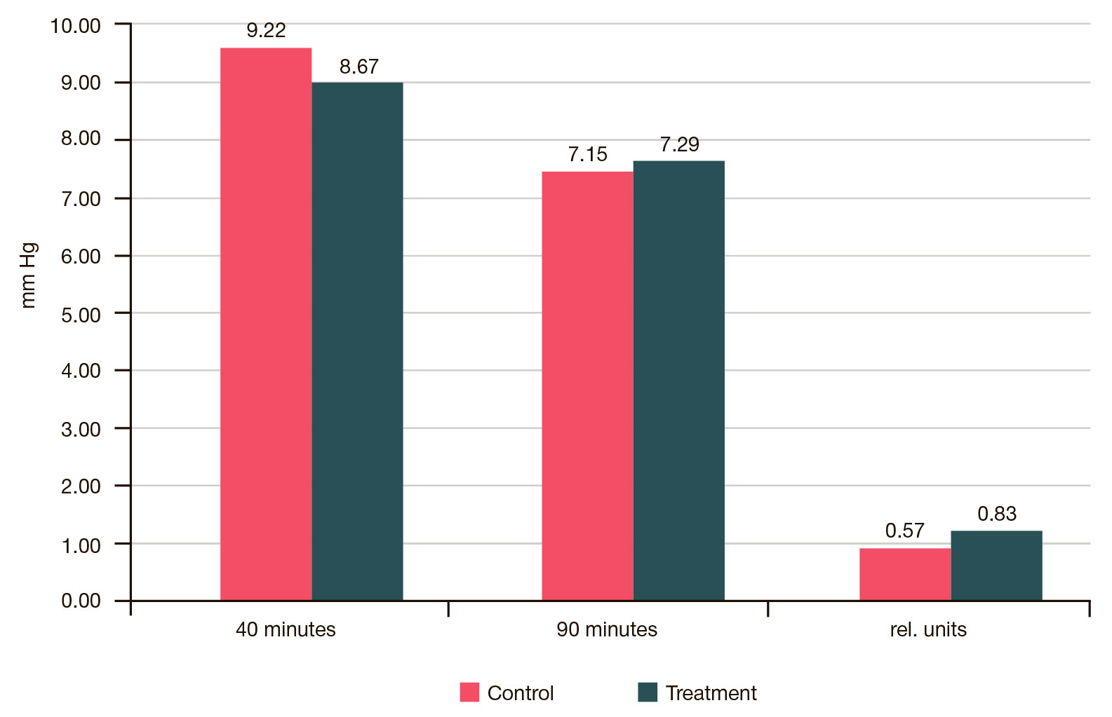
Fig. 4. Isolated lung perfusion pressure dynamics. The ratio of initial and final perfusion pressure is given in relative units
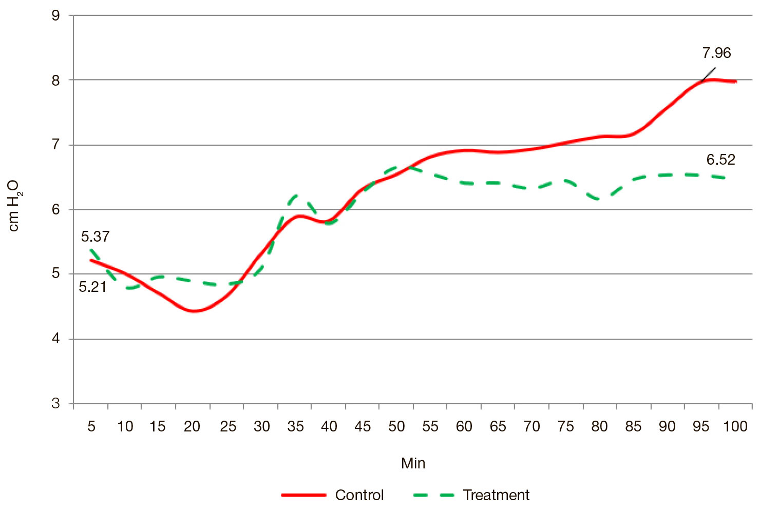
Fig. 5. Isolated lung intratracheal pressure dynamics
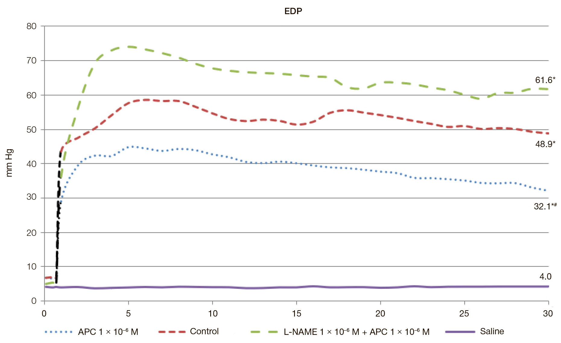
Fig. 6. End diastolic pressure dynamics, isolated heart during reperfusion after 30 minutes of total normothermic (37 °C) ischemia. APC administered. * — the differences are statistically significant in comparison with the intact organ; # — the differences are statistically significant in comparison with the "reperfusion (control)" group (р ˂ 0.05)
Table 1. Heart-lungs complex weight (M ± SD), n = 7
Note: * — statistically significant differences with the control series of experiments.
Table 2. Indicators of myocardial contractility with APC administered. The data provided are relative to the background, in % (M ± SE); for the EDP the data is absolute, n = 7
Note: * — the differences are statistically significant in comparison with the intact organ; # — the differences are statistically significant in comparison with the "reperfusion (control)" group (р ˂ 0.05)