ORIGINAL RESEARCH

Assessing hepatoprotective effects of antioxidants on amiodarone-induced cytotoxicity in human hepatoma HepaRG cell line

Filimonova KS, Rogovskaya NYu, Beltyukov PP, Babakov VN
About authors

Research Institute of Hygiene, Occupational Pathology and Human Ecology of the Federal Medical Biological Agency, Leningrad Region, Russia

Correspondence should be addressed: Vladimir Nikolaevich Babakov
Kapitolovo, 93, p/o Kuzmolovsky, Leningradskaja oblast, 188663; ur.hcepg@vokabab
Received: 2021-07-20 Accepted: 2021-08-11 Published online: 2021-09-23
|
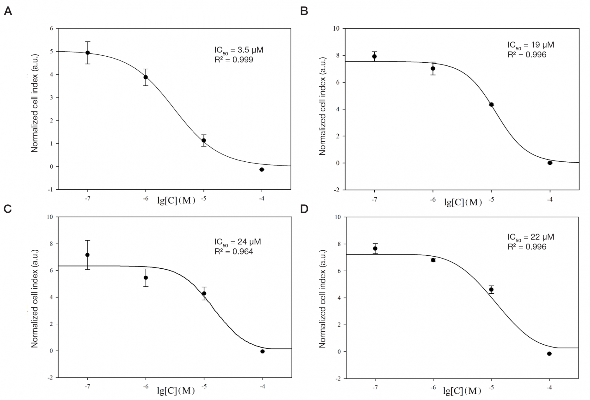
Fig. 1. Relationship between cell index of HepaRG cells and log concentration of amiodarone (cell index has been normalized in terms of the time when the medium containing the studied substance has been added). А. Changes in cell index upon exposure to amiodarone at a concentration of 0.1, 1, 10 and 100 μМ. B. Changes in cell index upon exposure to amiodarone dilution series in the presence of 100 μМ of vitamin Е. C. Changes in cell index upon exposure to amiodarone dilution series in the presence of 100 μМ of N-acetylcysteine. D. Changes in cell index upon exposure to amiodarone dilution series in the presence of 100 μМ of S- adenosylmethionine
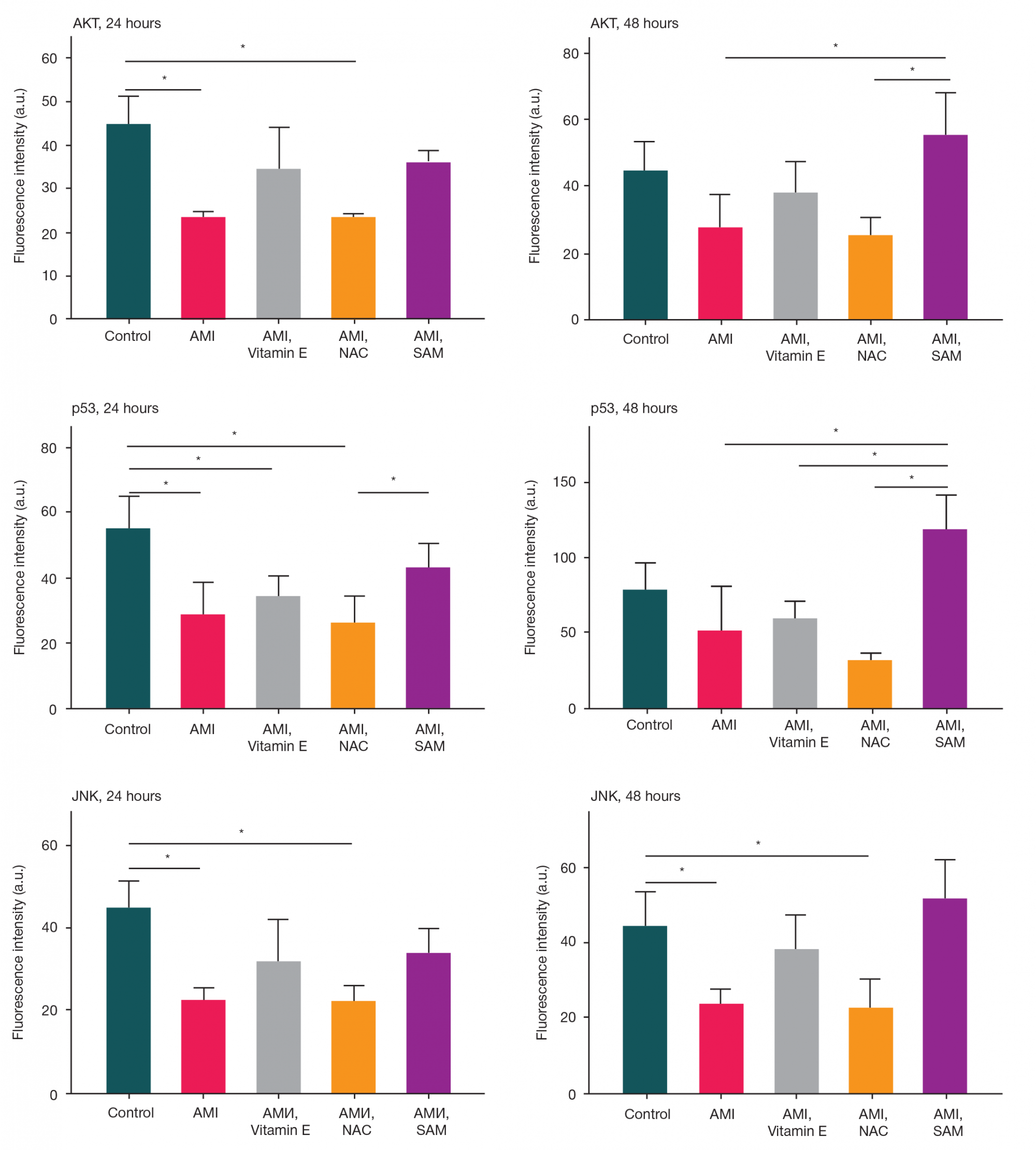
Fig. 2. Phosphorylated AKT kinase (Ser473), р53 protein (Ser46), JNK kinase (Thr183/Tyr185) fluorescence intensity in the HepaRG cell lysates after exposure to amiodarone at a concentration of 10 μМ (AMI), and against the effects of 100 μМ of vitamin Е, 100 μМ of N-acetylcysteine (NAC), 100 μМ of S-adenosylmethionine (SAM) during the 24- and 48-hour incubation. * — p < 0.05
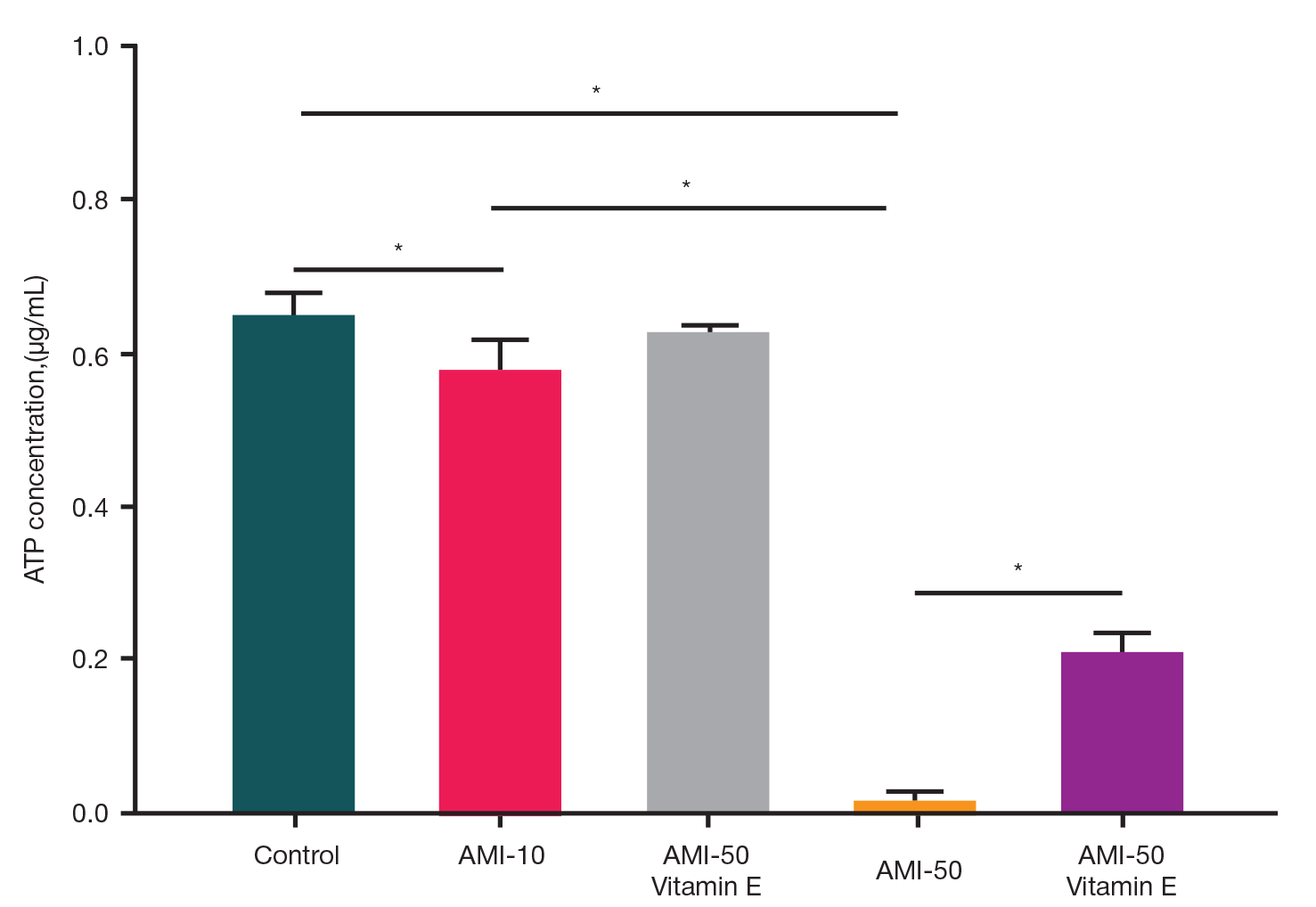
Fig. 3. ATP levels in HepaRG cell lysates after the 24-hour exposure to amiodarone at a concentration of 10 μМ (AMI-10) and 50 μМ (AMI-50) against the background of exposure to vitamin Е. * — p < 0.05
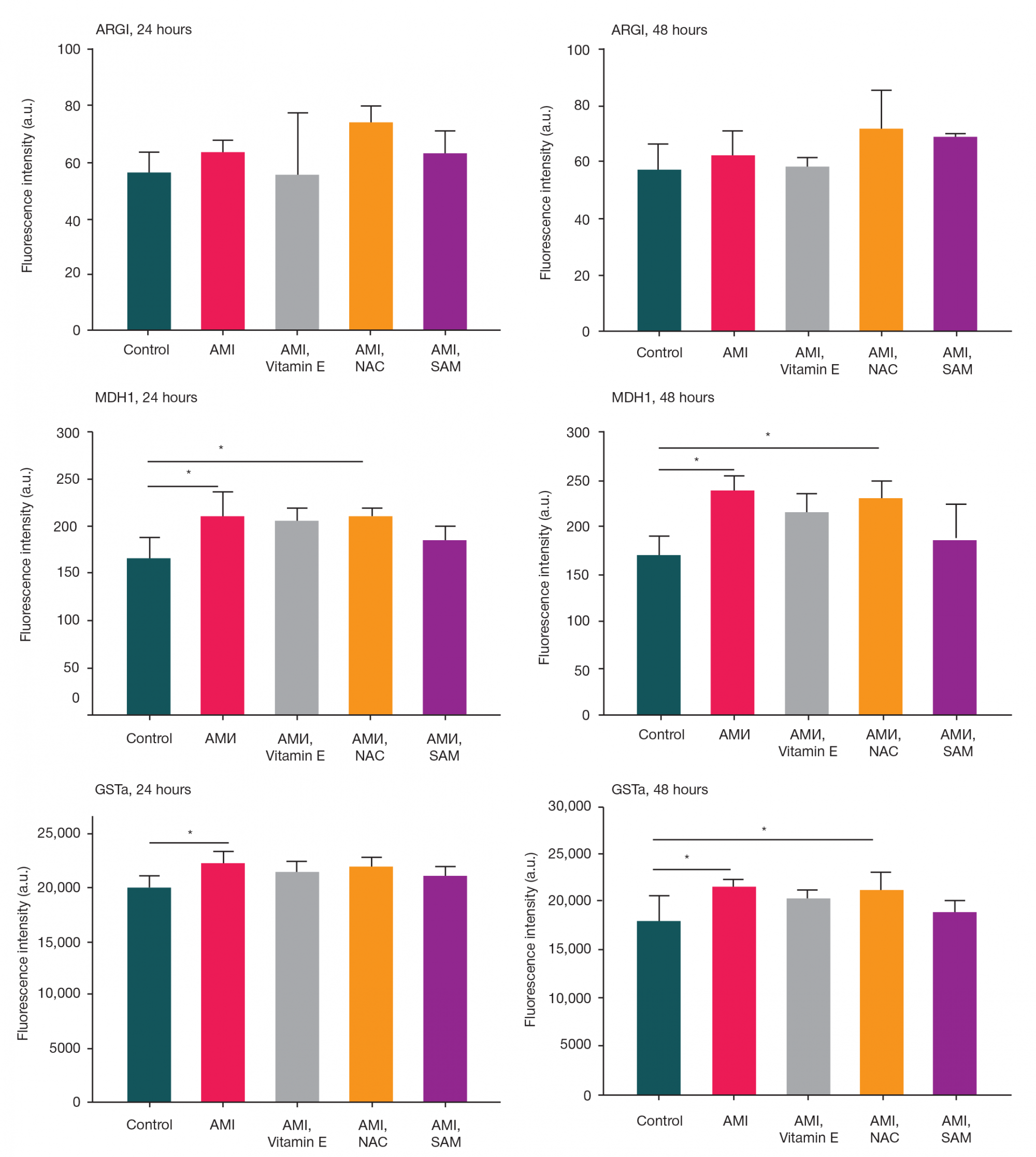
Fig. 4. Arginase 1 (ARG1), malate dehydrogenase 1 (MDH1), glutathione S-transferase (GSTα) fluorescence intensity in the conditioned medium of the HepaRG cells after exposure to amiodarone at a concentration of 10 μМ (AMI), and against the effects of 100 μМ of vitamin Е, 100 μМ of N-acetylcysteine (NAC), 100 μМ of S-adenosylmethionine (SAM) during the 24- and 48-hour incubation. * — p < 0.05
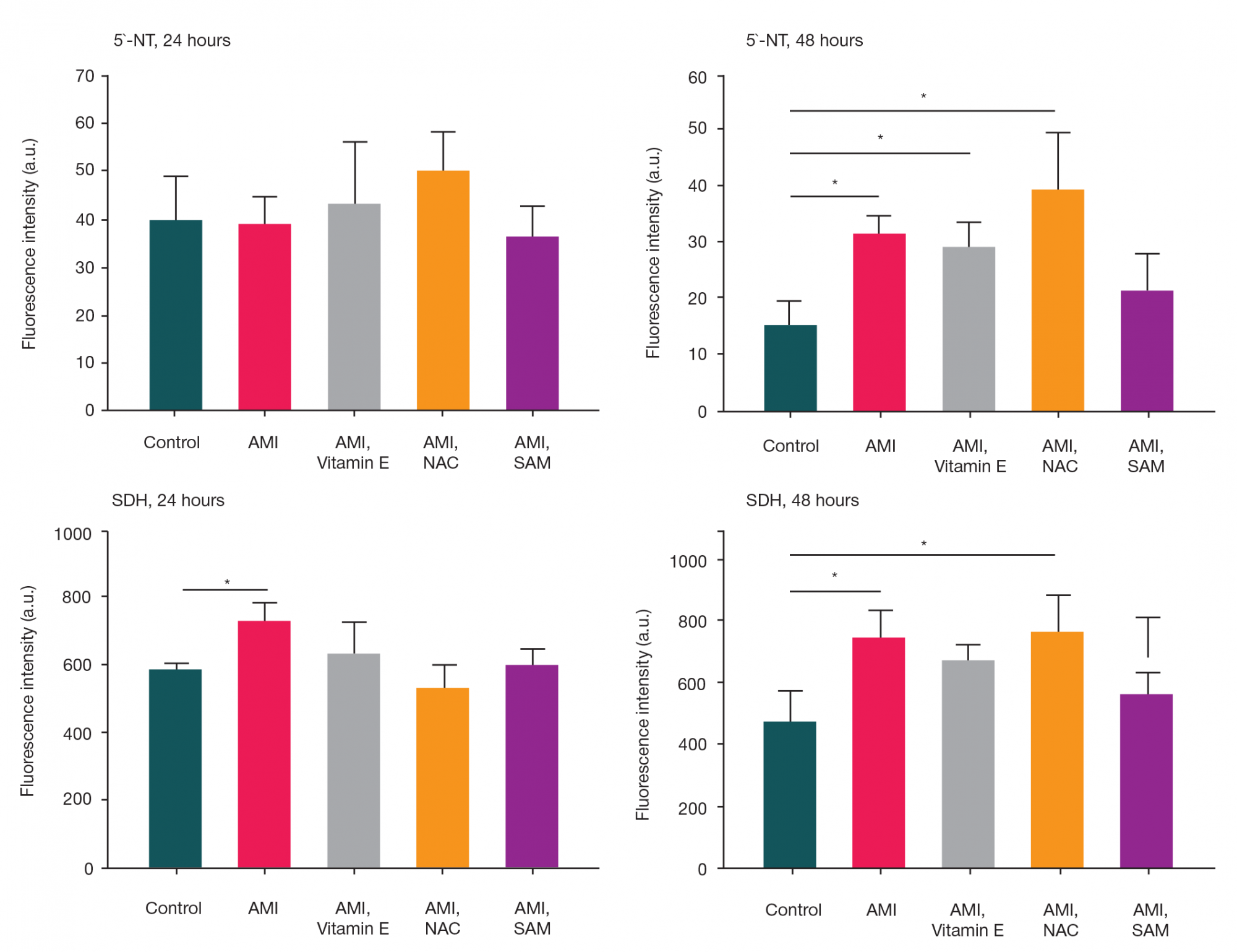
Fig. 5. Nucleotidase (5'-NT), sorbitol dehydrogenase (SDH) fluorescence intensity in the conditioned medium of the HepaRG cells after exposure to amiodarone at a concentration of 10 μМ (AMI), and against the effects of 100 μМ of vitamin Е, 100 μМ of N-acetylcysteine (NAC), 100 μМ of S-adenosylmethionine (SAM) during the 24- and 48-hour incubation. * — p < 0.05
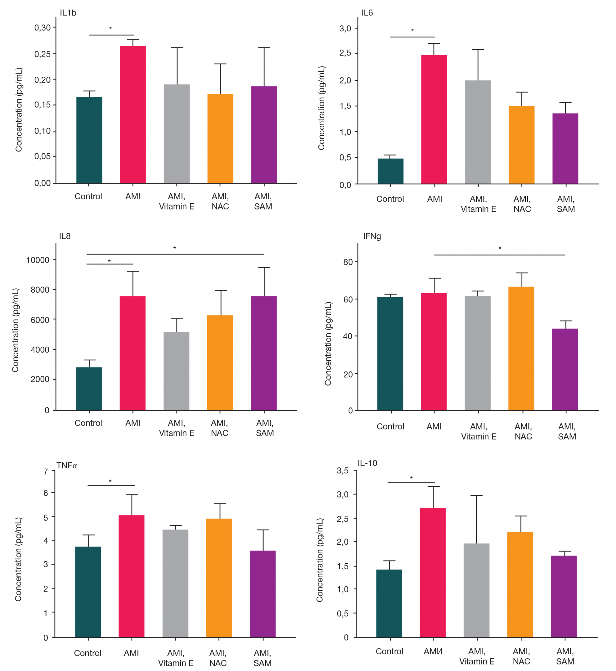
Fig. 6. Concentration of cytokines IL1β, IL6, IL8, IFNγ, TNFα and IL10 in the conditioned medium of HepaRG cells, incubated during 48 hours and exposed to amiodarone at a concentration of 10 μМ (AMI), and against the effects of 100 μМ of vitamin Е, 100 μМ of N-acetylcysteine (NAC), 100 μМ of S-adenosylmethionine (SAM). * — p < 0.05