Copyright: © 2021 by the authors. Licensee: Pirogov University.
This article is an open access article distributed under the terms and conditions of the Creative Commons Attribution license (CC BY).

CLINICAL CASE

Central vein sign for differential diagnosis of demyelinating diseases of CNS

About authors

1 Pirogov Russian National Research Medical University, Moscow, Russia

2 Federal Center for Brain Research and Neurotechnologies of FMBA, Moscow, Russia

Correspondence should be addressed: Alexey N. Boyko
Ostrovityanova, 1, 117437, Москва; moc.liamg@31naokyob

About paper

Author contribution: Belov SE — literature analysis, study design, recruitment of patients, clinical analysis, manuscript preparation; Gubsky IL — study design, MRI examinations, MRI data analysis, manuscript preparation; Lelyuk VG — study design, MRI data analysis, manuscript preparation; Boyko AN — study design, recruitment of patients, clinical analysis, manuscript preparation.

Compliance with ethical standards: informed consent was obtained from both patients.
Received: 2021-07-15 Accepted: 2021-08-04 Published online: 2021-08-18
|
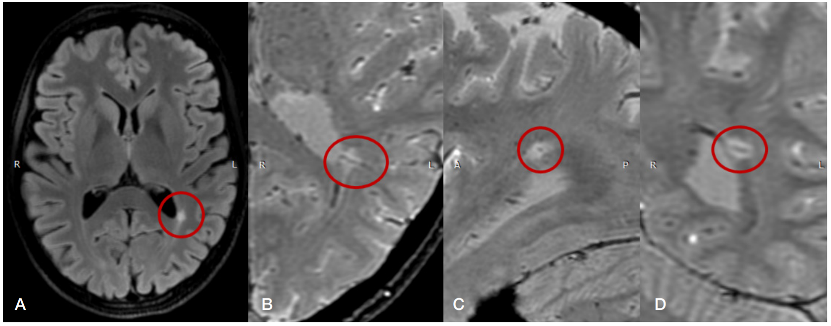
Fig. 1. Magnetic resonance images of patient 1. A. A T2-weighted fluid-attenuated inversion recovery (FLAIR) image, the axial plane. The image shows a periventricular lesion in the posterior horn of the left lateral ventricle (marked by the red oval). B. A susceptibility-weighted (SW) image, the axial plane. The image shows a central vein sign in the lesion near the posterior horn of the left ventricle (marked by the red oval). C. A susceptibility-weighted (SW) image; the sagittal plane, the central vein sign appears as a dot in the lesion (marked by the red oval). D. A susceptibility-weighted (SW) image, the coronal plane; the central vein sign appears as a thin line in the same lesion (marked by the red oval)
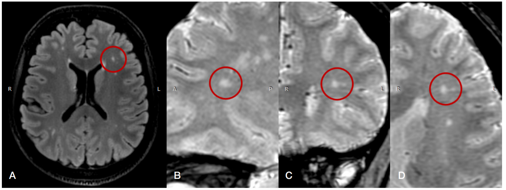
Fig. 2. Magnetic resonance images of patient 2. A. A T2-weighted fluid-attenuated inversion recovery (FLAIR) image, the axial plane. The image shows a round lesion in the deep white matter of the left frontal lobe (marked by the red oval). B. A susceptibility-weighted (SW) image, the axial plane. The image shows the same lesion, the central vein sign is not visualized (the red oval). C. A susceptibility-weighted (SW) image, the sagittal plane. The same lesion, the central vein sign is not visualized; a smaller lesion not suitable for the analysis is located in close proximity (the red oval). D. A susceptibility-weighted (SW) image, the coronal plane. The same lesion, the central vein sign is not visualized (the red oval)