ORIGINAL RESEARCH

Interaction of cationic antiseptics with cardiolipin-containing model bacterial membranes

Kholina EG1,2, Bozdaganyan ME1,2, Strakhovskaya MG1,2, Kovalenko IB1,2
About authors

1 Federal Scientific and Clinical Center of Specialized Medical Care and Medical Technology of FMBA, Moscow, Russia

2 Lomonosov Moscow State University, Moscow, Russia

Correspondence should be addressed: Ilya B. Kovalenko
Orekhovyi bulvar, 28, Moscow, 115682; moc.liamg@87oknelavoki

About paper

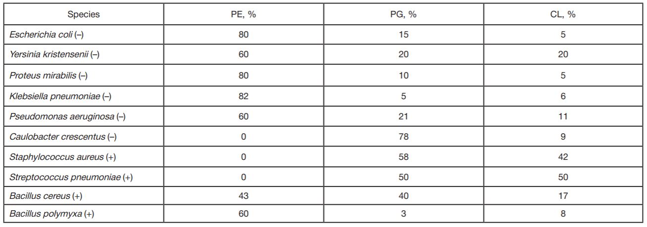
Funding: the research was carried out with the financial support of the Russian Foundation for Basic Research (project № 19-34-90045) and the State assignment "The influence of the lipid composition of bacterial membranes on the processes of interaction with antimicrobial compounds" (code: "Membrane").

Author contribution: Kholina EG — constructing molecular models of studied substances, calculations, manuscript writing; Bozdaganyan ME — calculations, manuscript writing; Strakhovskaya MG — study concept, manuscript writing, analysis of the results; Kovalenko IB — study concept, building a computing infrastructure, manuscript writing, analysis of the results.

Received: 2021-07-19 Accepted: 2021-08-21 Published online: 2021-09-11
|
Fig. 1. CA chemical structures with overlapping coarse grains. А. Miramistin (MIR). B. Сhlorhexidine (CHL). C. Picloxidine (PIC). D. Octenidine (OCT). Coarse grains are highlighted in different colors in accordance with the particle type selected in MARTINI force field (bottom part of A panel)
Fig. 2. Characteristics of model membrane comprising POPE:POPG:CDL2 in the presence of various CA concentrations. А. Lateral diffusion coefficients for the following lipids: POPE (left), POPG (centre) and СDL2 (right); B. Bilayer thickness; C. Area per lipid. (Parameter values for model membrane obtained without adding CAs are marked in gray)
Fig. 3. Effect of CDL2 clustering in the presence of antiseptic OCT at the CA : lipid ratio of 1 : 4. А. Image of MD calculation at time 15 µs. POPE lipids are marked in green, POPG in pink, CDL2 in red, and OCT in blue. Phosphate residues are marked in black, and charged particles of OCT molecules are marked in purple. B. Density profiles for various components of model membrane. Density profile for lipid fatty acid chains is marked in pink, charged particles of OCT are marked in cyan, terminal OCT particles are marked in light blue, NH3 particle (ethanol) of POPE lipid is marked in purple, GL0 particle (glycerol) of POPG lipid is marked in green. Position of phosphates is represented by the dotted lines going through the centers of corresponding peaks. Density profiles for fatty acid chains normalized to the maximum peak value are shown on the second Y axis (right)
Fig. 4. Effect of pulling lipids out of the model bilayer by OCT molecules. CA : lipid ratio of 1 : 4. А. Consecutive images of MD calculation obtained at different times. POPE lipids are marked in green, POPG in pink, CDL2 in red, and OCT in blue. Phosphate residues are marked in black, and charged particles of OCT molecules are marked in purple. B. Number of lipids pulled out of the bilayer as a function on MD calculation time
Table. Ratios of phosphatidylethanolamine (PE), phosphatidylglycerol (PG), cardiolipin (CL) in plasma membranes of some species of gram-negative (–) and gram-positive (+) bacteria [13, 14]