CLINICAL CASE

Focal laser photocoagulation of the optic disc peripapillary neovascularization in patient with proliferative diabetic retinopathy

About authors

Pirogov Russian National Research Medical University, Moscow, Russia

Correspondence should be addressed: Ekaterina P. Tebina
Volokolamskoe shosse, 30, korp. 2, Moscow, 123182, Russia; ur.liam@anibetaniretake

About paper

Author contribution: Takhchidi KhP — study concept and design, manuscript editing; Takhchidi NKh — literature analysis; Tebina EP — data acquisition and processing, manuscript writing; Kasminina TA — laser therapy.

Compliance with ethical standards: the patient submitted the informed consent to laser therapy and personal data processing.
Received: 2022-01-15 Accepted: 2022-03-01 Published online: 2022-03-13
|
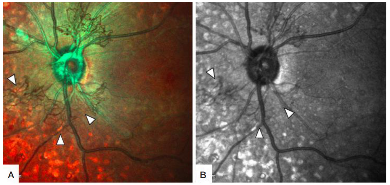
Fig. 1. Prior to laser therapy. А. Multispectral image; B. Infrared scanning laser ophthalmoscopy: network of the newly formed blood vessels is visible primarily in the upper nasal, lower nasal, and lower temporal sectors of the peripapillary region (white arrows)
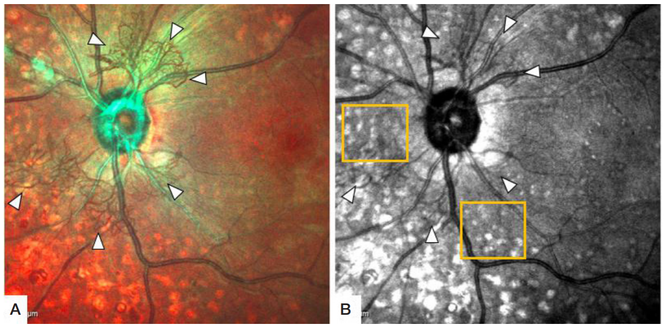
Fig. 2. A week after the first stage of laser photocoagulation. А. Multispectral image: network of the newly formed blood vessels in the peripapillary region is still active (white arrows). B. Infrared scanning laser ophthalmoscopy: network of the newly formed blood vessels in the peripapillary region is still active (white arrows), fresh coagula are observed in the peripapillary region (yellow square)
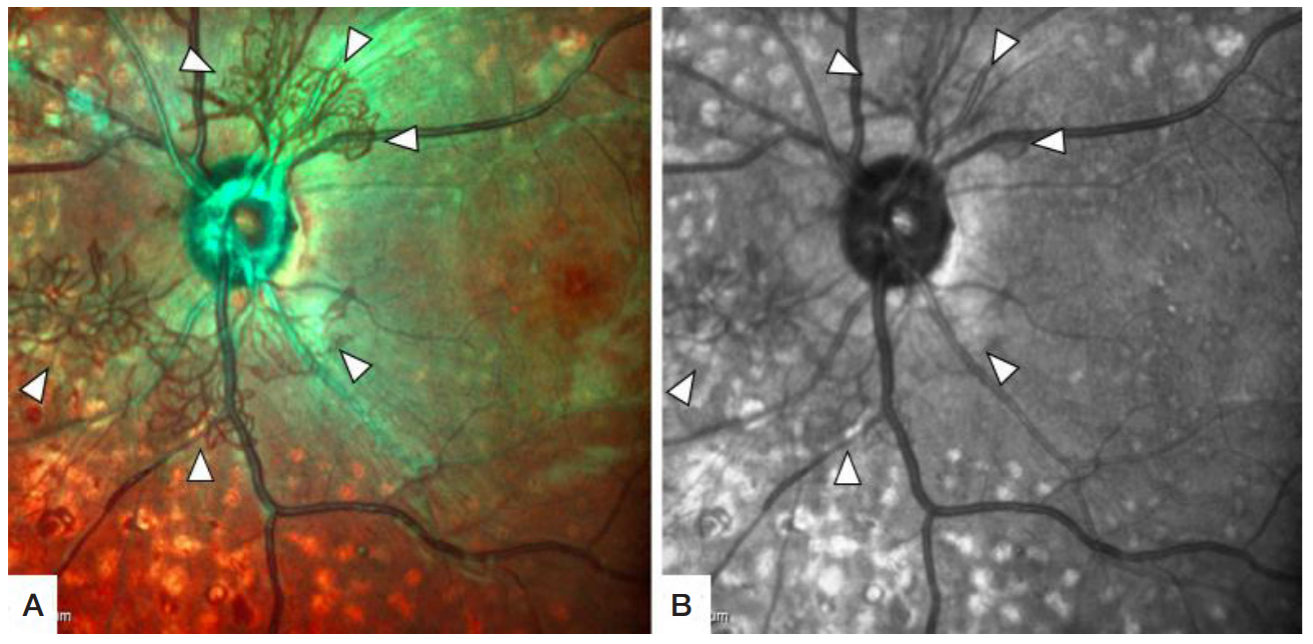
Fig. 3. Four months after the second-stage laser photocoagulation of the newly formed blood vessels. А. Multispectral image. B. Infrared scanning laser ophthalmoscopy: emptied newly formed vessels are visible in the peripapillary region (white arrows)
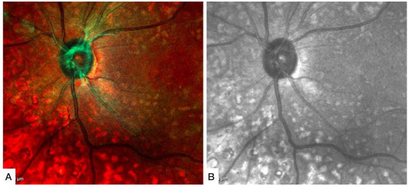
Fig. 4. Four years after the second-stage laser photocoagulation of the newly formed blood vessels. А. Multispectral image. B. Infrared scanning laser ophthalmoscopy: complete regression of the optic disc network of the newly formed vessels is observed in the peripapillary region