ORIGINAL RESEARCH

Neurophysiological assessment of speech function in individuals having a history of mild COVID-19

Gulyaev SA, Voronkova YuA, Abramova TA, Kovrazhkina EA
About authors

Federal Center of Brain Research and Neurotechnologies of the Federal Medical Biological Agency, Moscow, Russia

Correspondence should be addressed: Sergey A. Gulyaev
Ostrovitianova, 1, str. 10, Moscow, 117997, Russia; ur.xednay@ssurgres

About paper

Author contribution: Gulyaev SA — data analysis, manuscript writing, editing; Voronkova YuA, Abramova TA — data acquisition; Kovrazhkina EA — editing.

Compliance with ethical standards: the study was approved by the Ethics Committee of the Federal Center for Brain and Neurotechnologies of FMBA (protocol № 148-1 dated June 15, 2021). All the subjects took part in the experiment on a voluntary basis with no extra benefit. The experiment was studied by employees of the Federal Center for Brain and Neurotechnologies of FMBA within the limits of scientific work conducted by the institution with no third party funding.
Received: 2022-03-24 Accepted: 2022-05-04 Published online: 2022-05-25
|
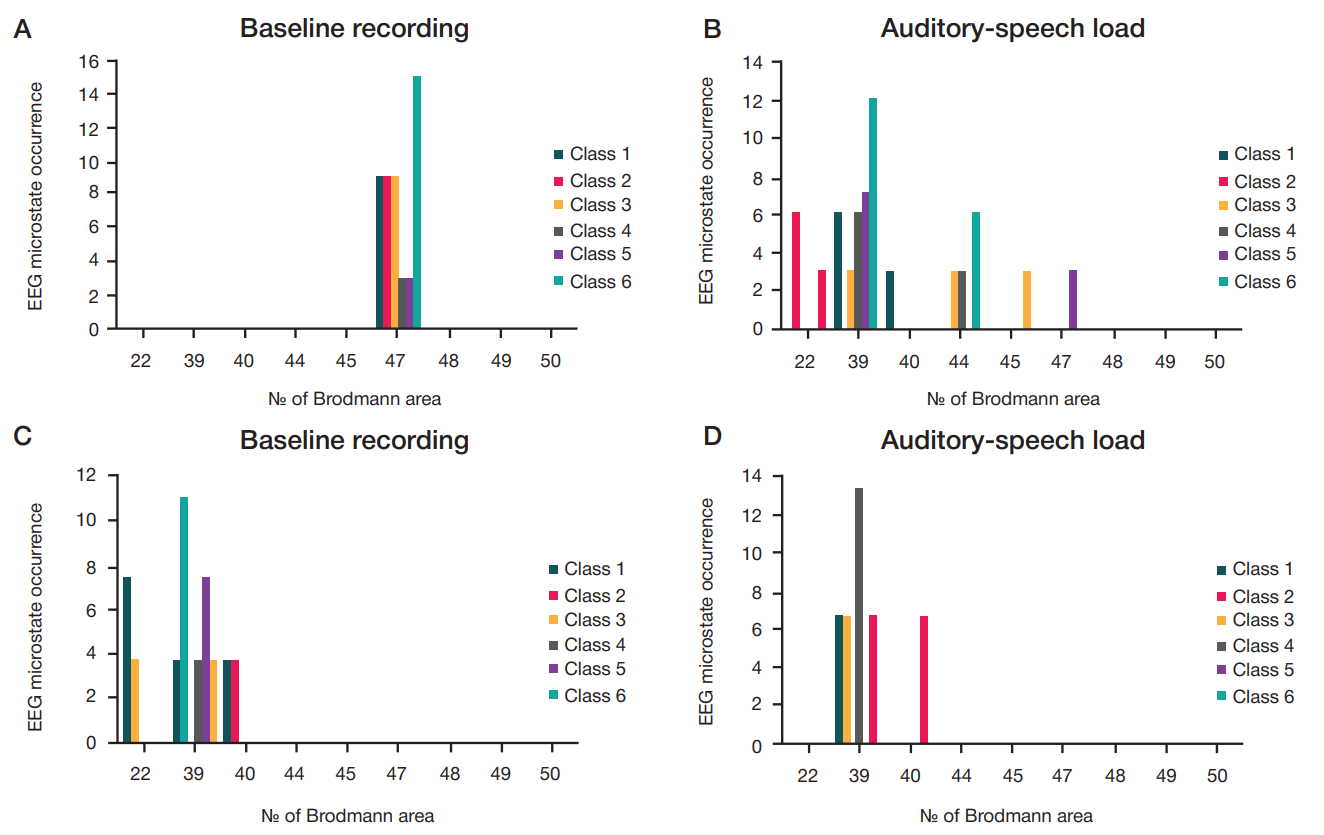
Fig. Rhythmic activity occurrence in accordance with the Brodmann area in controls (А, B) and individuals having a history of novel coronavirus infection (C, D) in the state of relaxed wakefulness and under auditory-speech load; p < 0.05 (χ2-test)
Table 1. Comparative characteristics of the class 1–6 EEG microstate duration in controls
Note: m — mean; SD — standard deviation.
Table 2. Comparative characteristics of the class 1–6 EEG microstate duration in individuals having a history of novel coronavirus infection
Notе: m — mean; SD — standard deviation.
Table 3. Comparative characteristics of the class 1–6 EEG microstate occurrence in controls
Notе: m — mean; SD — standard deviation.
Table 4. Comparative characteristics of the class 1–6 EEG microstate occurrence in individuals having a history of novel coronavirus infection
Notе: m — mean; SD — standard deviation.
Table 5. Comparative characteristics of the class 1–6 EEG microstate coverage in controls
Notе: m — mean; SD — standard deviation.
Table 6. Comparative characteristics of the class 1–6 EEG microstate coverage in individuals having a history of novel coronavirus infection
Notе: m — mean; SD — standard deviation.