ORIGINAL RESEARCH

Transcription factors in human skeletal muscle associated with single and regular strength exercises

Lednev EM1,2, Makhnovskii PA2, Vepkhvadze TF2, Sultanov RI1, Zhelankin AV1, Kanygina AV1, Popov DV1,2, Generozov EV1
About authors

1 Lopukhin Federal Research and Clinical Center of Physical-Chemical Medicine of the Federal Medical Biological Agency, Moscow, Russia

2 Institute for Biomedical Problems of the Russian Academy of Sciences, Moscow, Russia

Correspondence should be addressed: Egor M. Lednev
Khoroshevskoe shosse, 76A, Moscow, 123007, Russia moc.liamg@zuahdel

About paper

Funding: the study was supported financially by the Russian Science Foundation, Agreement № 21-15-00362 "Investigation of molecular genetic mechanisms of morphofunctional changes in human muscle fibers during high-intensity physical loads".

Author contribution: Lednev EM, Vepkhvadze TF — study design and conduct, muscle sampling; Makhnovskii PA, Sultanov RI and Kanygina AV — bioinformatic data analysis; Zhelankin AV, Lednev EM — laboratory research; Generozov EV, Popov DV — study design and conduct, data processing, article authoring.

Compliance with ethical standards: the study was approved by the Ethics Committee of the Lopukhin Federal Research and Clinical Center Of Physical-Chemical Medicine (Minutes № 202/06/01 of June 01, 2021). All participants signed the voluntary informed consent form.
Received: 2023-07-21 Accepted: 2023-09-01 Published online: 2023-09-27
|
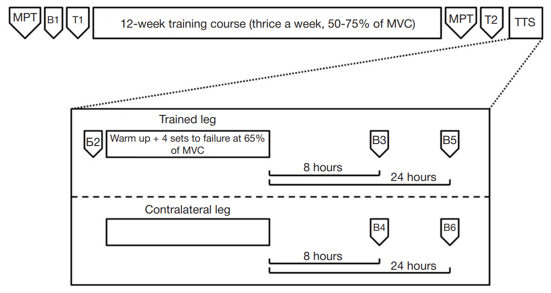
Fig. 1. Physiological experiment diagram. Т1, Т2 — testing the maximum voluntary contraction (MVC); TTS — test training session (single session with bioptic samples taken from both legs before and after exercising one leg); B1–B6 — sampling from the m. vastus lateralis. Biopsies B1, B2, B3, B5 were taken from the leg loaded during TTS. Biopsies B4 and B6 — from the contralateral leg (which was not loaded). Both legs were exercised during twelve-week training course
Fig. 2. Number of genes that changed expression in m. vastus lateralis after a 12-week training program and 8 and 24 hours after a single load. Venn diagrams show the number of mRNAs unique and common to different experimental conditions for genes that increased and decreased expression
Fig. 3. Transcription factors (TFs) associated with genes that increased and decreased expression after regular strength training sessions (Training) and 8 hours and 24 hours after a single physical load. Shades of color and numbers indicate the amount of enrichment of the binding motif with the transcription factor in individual promoters of genes that have changed expression, relative to 5000 random genes that have not changed expression (see METHODS).
Table 1. Results of analysis of functional enrichment, genes that changed expression in m. vastus lateralis after a training course and a one-time physical load