REVIEW

Morphological characteristics of toxic brain damage

Gaykova ON1, Kozlov AA1, Katretskaya GG1, Melnikova MV1, Melekhova AS1, Bondarenko AA1, Sokolova YuO1, Bazhanova ED1,2
About authors

1 Golikov Research Clinical Center of Toxicology of the Federal Medical and Biological Agency, Saint-Petersburg, Russia

2 Sechenov Institute of Evolutionary Physiology and Biochemistry of the Russian Academy of Sciences, Saint-Petersburg, Russia

Correspondence should be addressed: Elena D. Bazhanova
Bekhtereva, 1, Saint-Petersburg, Russia; 192019; ur.liam@eavonahzab

About paper

Funding: the study was conducted as part of the State Assignment of the FMBA of Russia No. 388-00071-24-00 (theme code 64.004.24.800) and supported by the State Assignment of the Sechenov Institute of Evolutionary Physiology and Biochemistry RAS No. 075-00264-24-00.

Author contribution: Gaikova ON, Sokolova YuO, Bazhanova ED, Kozlov AA – literature review, data acquisition and processing, manuscript writing and editing; Katretskaya GG, Melnikova MV, Melekhova AS, Bondarenko AA – editing, formatting, approval of the final version of the article.
Received: 2024-05-15 Accepted: 2024-06-17 Published online: 2024-06-28
|
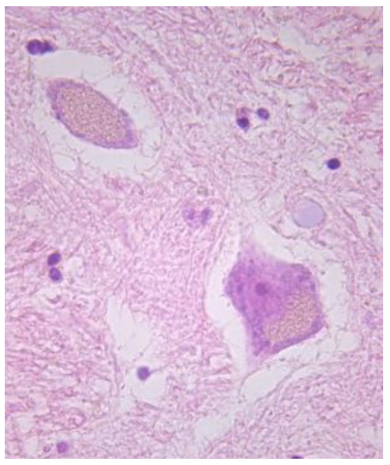
Fig. 1. Lipofuscin in the cytoplasm of neurons, multiple light-yellow granules, sectional material. Hematoxylin and eosin staining, ×630
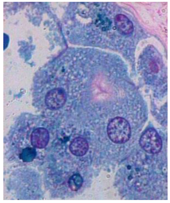
Fig. 2. Lipofuscin in the epithelium of the vascular plexus, blue-green granules in the cytoplasm, sectional material. Staining with alcian blue, ×1000
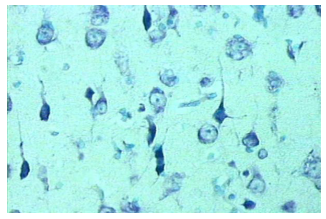
Fig. 3. Temporal lobe cortex acute swelling of neurons, shadow cells, dark, shriveled neurons, biopsy material. Nissl staining, ×400
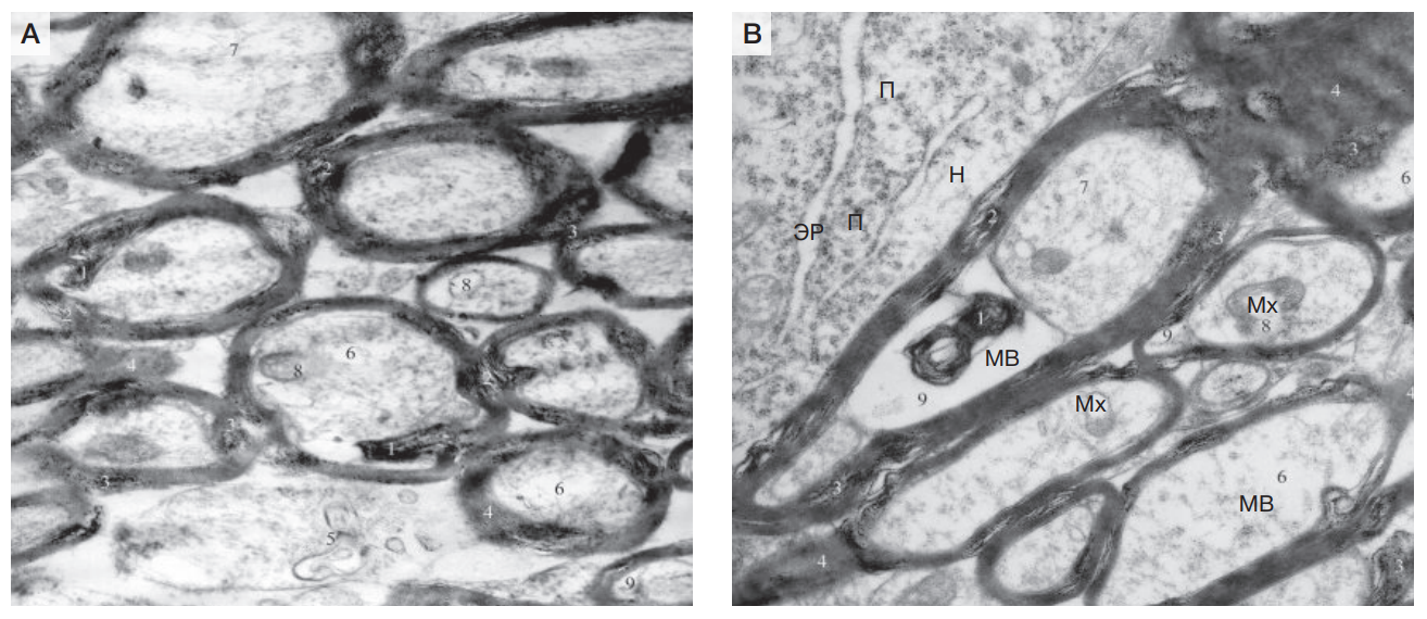
Fig. 4. Changes in myelinated fibers of the white matter Fig. 4A — ×20,000.; Fig. 4B — ×16000. MF — myelin fiber; N — neuron; ER — endoplasmic reticulum; P —polysomes; Mch — mitochondrion; 1 — Disruption of myelin sheath configuration; 2 — delamination; 3 — granular disintegration; 4 — homogenization; 5 — demyelination; 6 — brightening of the axial cylinder matrix; 7— disorientation of neurofilaments; 8 — destruction of mitochondria; 9 — periaxonal edema, experimental material (rats). Electronograms
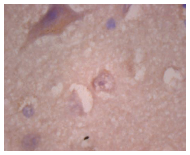
Fig. 5. Oncosuppressor gene is determined in the nucleus of the "dementia neuron". The cytoplasm of a large neuron of typical structure has a large amount of lipofuscin (sectional material). Immunohistochemical reaction of p53, ×1000
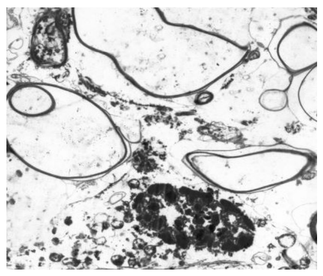
Fig. 6. Sections of white matter. Filamentous myelin fibers with transparent axial cylinders. Near oligodendrocyte with signs of apoptosis (sectional material). Electronogram, ×5000
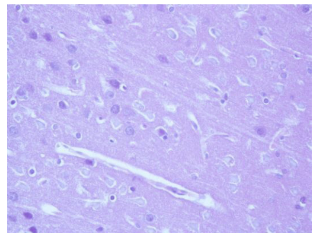
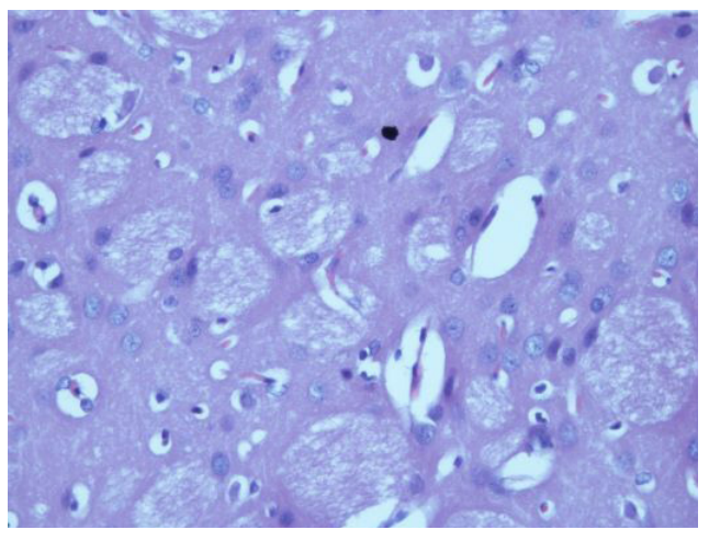
Fig. 7. Cerebral cortex. The perivascular spaces are dilated. Most neurons are "shadow cells", single neurons with neuronophagy phenomena. Foci of neuron loss, experimental material (rats). Hematoxylin and eosin staining, ×400
Fig. 8. Brain, subcortical nuclei. The perivascular spaces are significantly enlarged. Most neurons are unchanged, marked rarefaction of the neuropil of the conducting pathways, experimental material (rats). Hematoxylin and eosin staining, ×400