CLINICAL CASE

Simultaneous repair of the skull base and the frontal lobe defect using CAD-CAM technology

Ivanov OV
About authors

Federal Siberian Research and Clinical Center of FMBA, Krasnoyarsk, Russia

Correspondence should be addressed: Evgeniya A. Blinova
Vorovskogo, 68, korp. 1, Chelyabinsk, 454141, Russia; ur.mrcru@avonilb

About paper

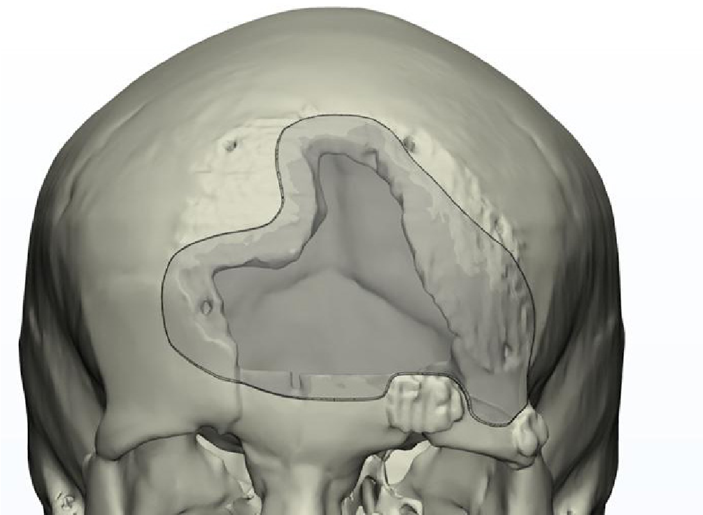
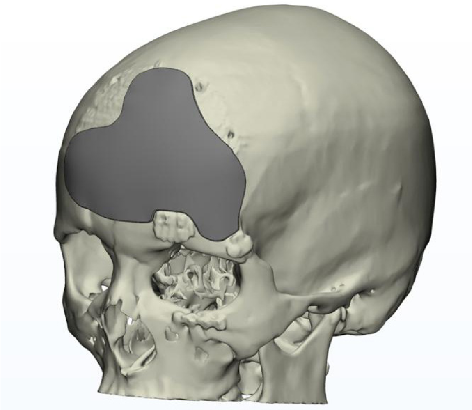
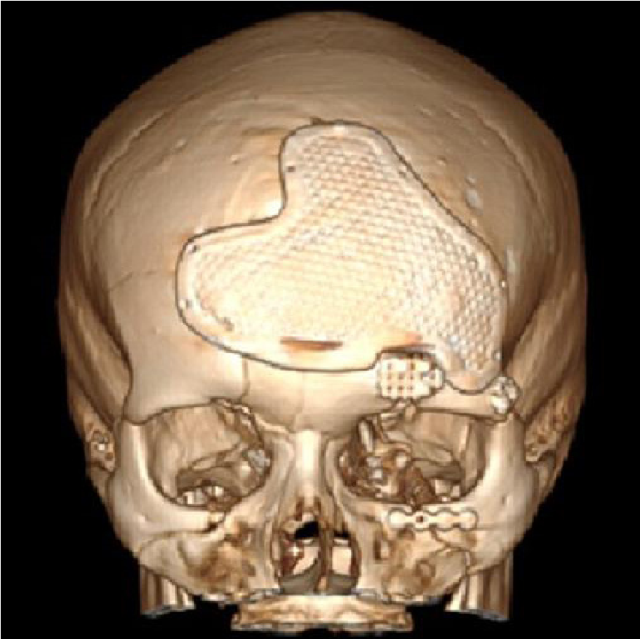
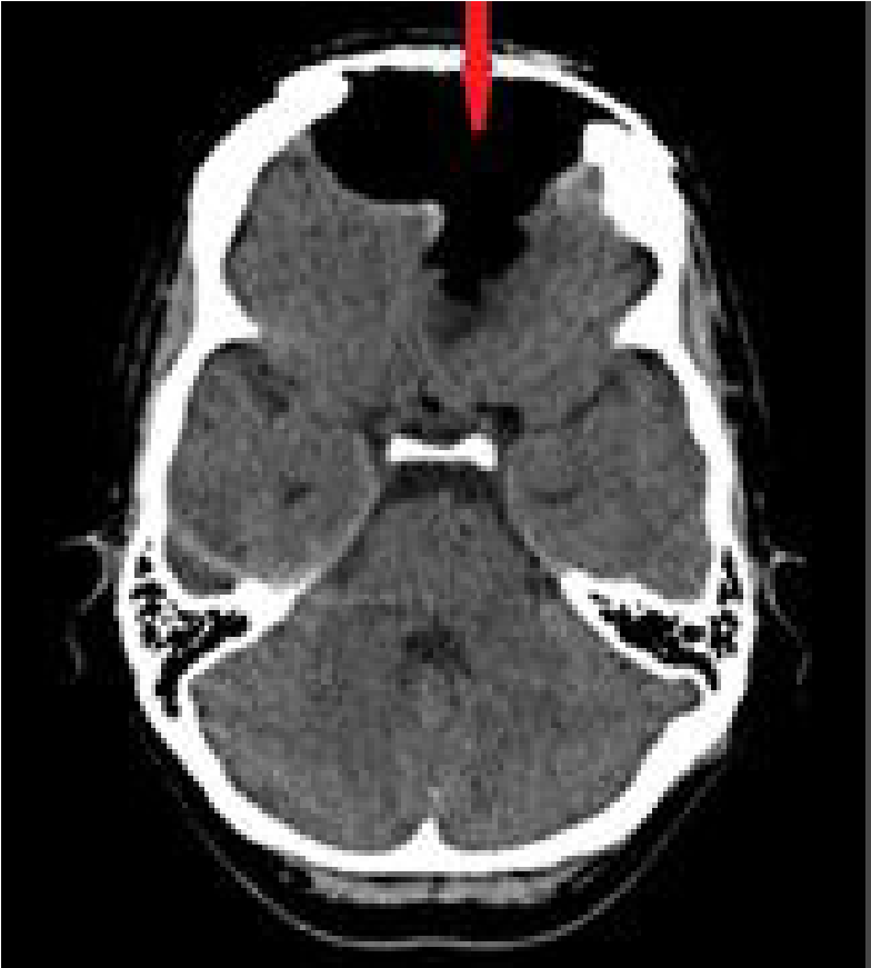
Funding: the work was part of the State Assignment on the Cranial bone defect repair with shape memory materials.

Acknowledgments: the authors thank Urasovsky IB, Director of Logeeks Medical Systems, and Panchencko AA, Director of Engineering, for their help in creating the customized implant.

Compliance with ethical standards: the patient gave her informed consent to participate in the study

Received: 2021-10-07 Accepted: 2021-11-08 Published online: 2021-11-08
|
Fig. 1. Computed tomography: 3D skull reconstruction, frontal view. A titanium mesh is visualized in the projection of the frontal bone defect. The image shows frontal lobe fragments forming the superior orbital wall after osteosynthesis
Fig. 2. Computed tomography: 3D skull reconstruction (side view)
Fig. 3. An axial CT image. The arrow points to the edge of the plate that deforms the cranial contour. The image shows the difference in geometry between the cranial vault and the implant
Fig. 4. An axial CT cisternography image
Fig. 5. 3D reconstruction of the skull: CAD planning. The implant is larger than the defect size, which is necessary for more accurate restoration of the frontal bone shape at the planning stage
Fig. 6. 3D reconstruction of the skull, CAD planning. The final version of the implant
Fig. 7. 3D reconstruction of the skull after the surgery (frontal view)
Fig. 8. The axial CT image after the surgical intervention
Fig. 9. Postoperative follow-up CT images