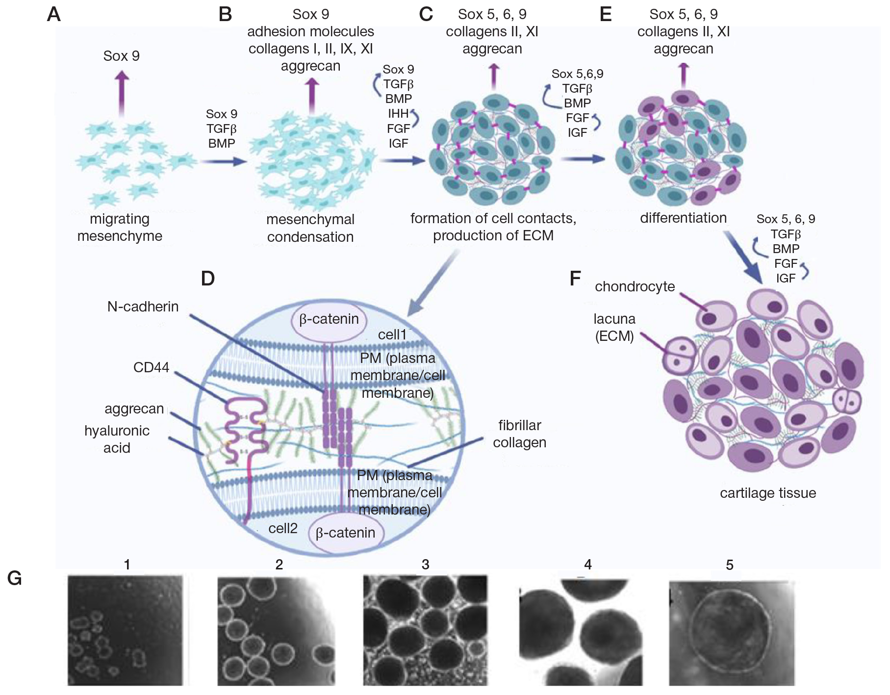
Fig. 1. General sequence of chondrogenesis processes. A. Migration of the mesenchyme. B. Prechondrogenic mesenchymal condensation. C. Formation of cell contacts, synthesis of ECM. D. Intercellular space and microenvironment of differentiating cells. E. Beginning of chondrogenic differentiation. F. Cartilage formation. G. Phase contrast microscopy pictures, magnification x10: 1-5 — spheroids after 1, 2, 3, 4 and 5 weeks of cultivation, 3D cultivation condition. ×100
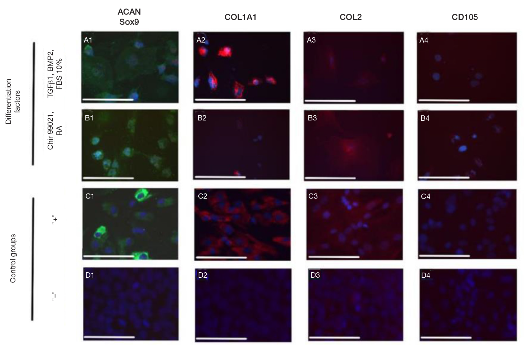
Fig. 2. Immunocytochemical analysis of monolayer cultures. A. Use of TGFβ1, BMP2 and 10% FBS. B. Using Chir99021 and RA. C. Articular chondrocyte culture, positive control. E. FD4S iPSC culture, negative control. 1 — aggrecan (green) and Sox9 (red), 2 — type I collagen (red), 3 — type II collagen (red), 4 — CD105 (red). Scale bar — 200 microns
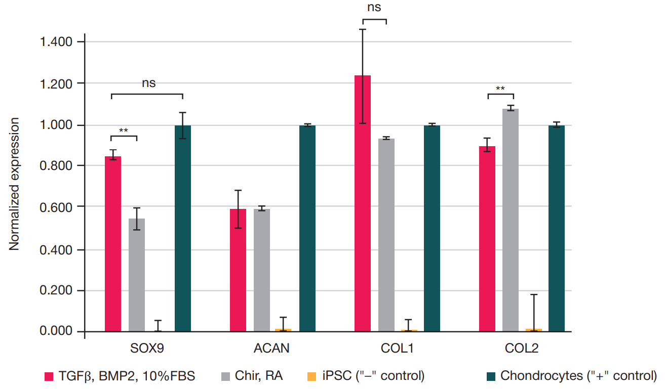
Fig. 3. Indicators of gene expression of chondrogenic markers in monolayer cultures. Error bar is the standard deviation. Significance of differences between groups: ns, p > 0.05; * — p < 0.05; * * — p < 0.01; * * * — p < 0.001
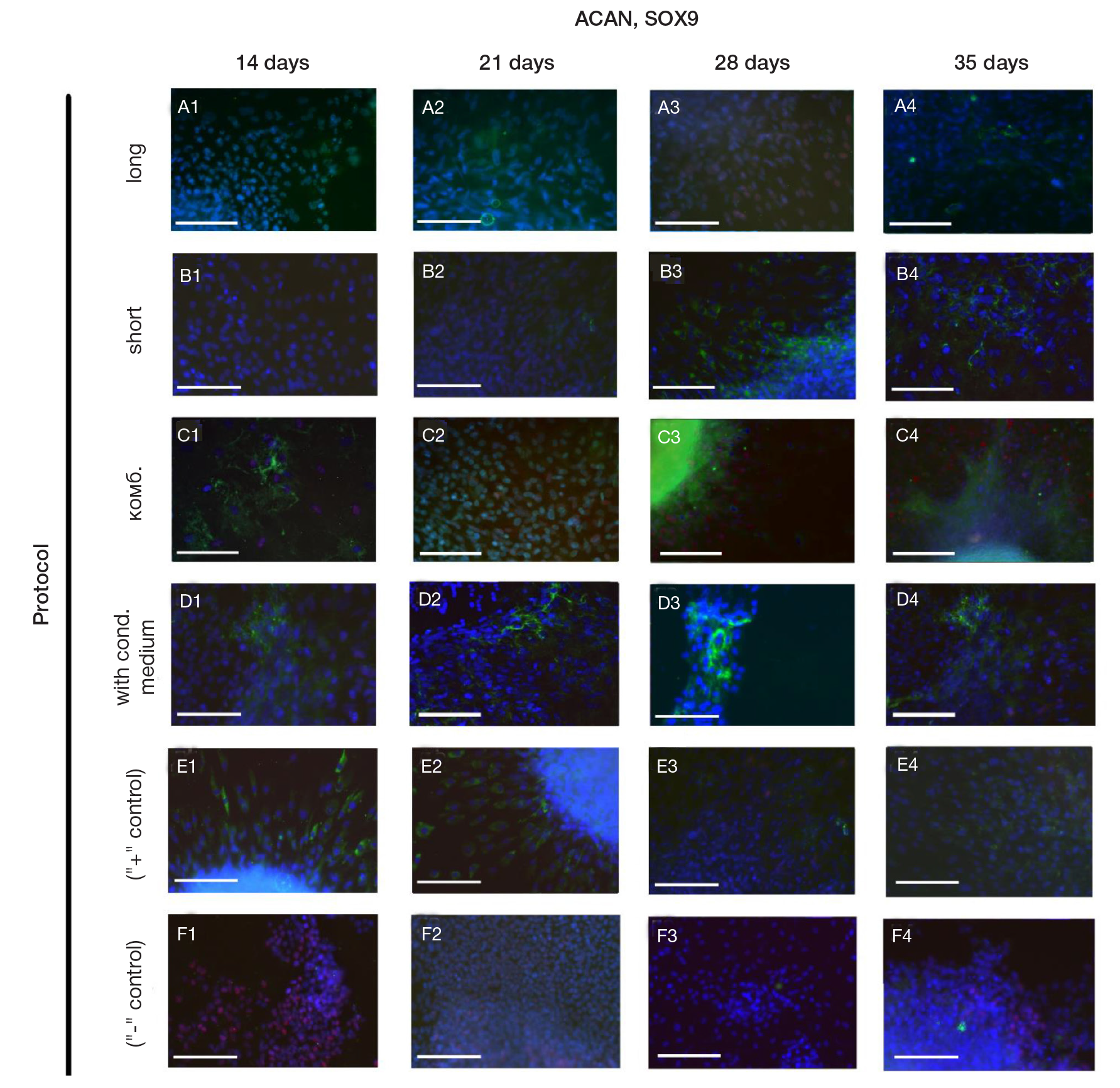
Fig. 4. Immunocytochemical analysis of aggrecan (green) and Sox9 (red) synthesis in 3D spheroid cultures of different protocols. A–D. Differentiation protocols: "long" (A), "short" (B), "combined" (C), with conditioned medium (D). E. Spheroids of the positive control group. F. Spheroids of the negative control group. Duration of differentiation: 1 — 14 days, 2 — 21 days, 3 — 28 days, 4 — 35 days. Scale bar — 200 microns
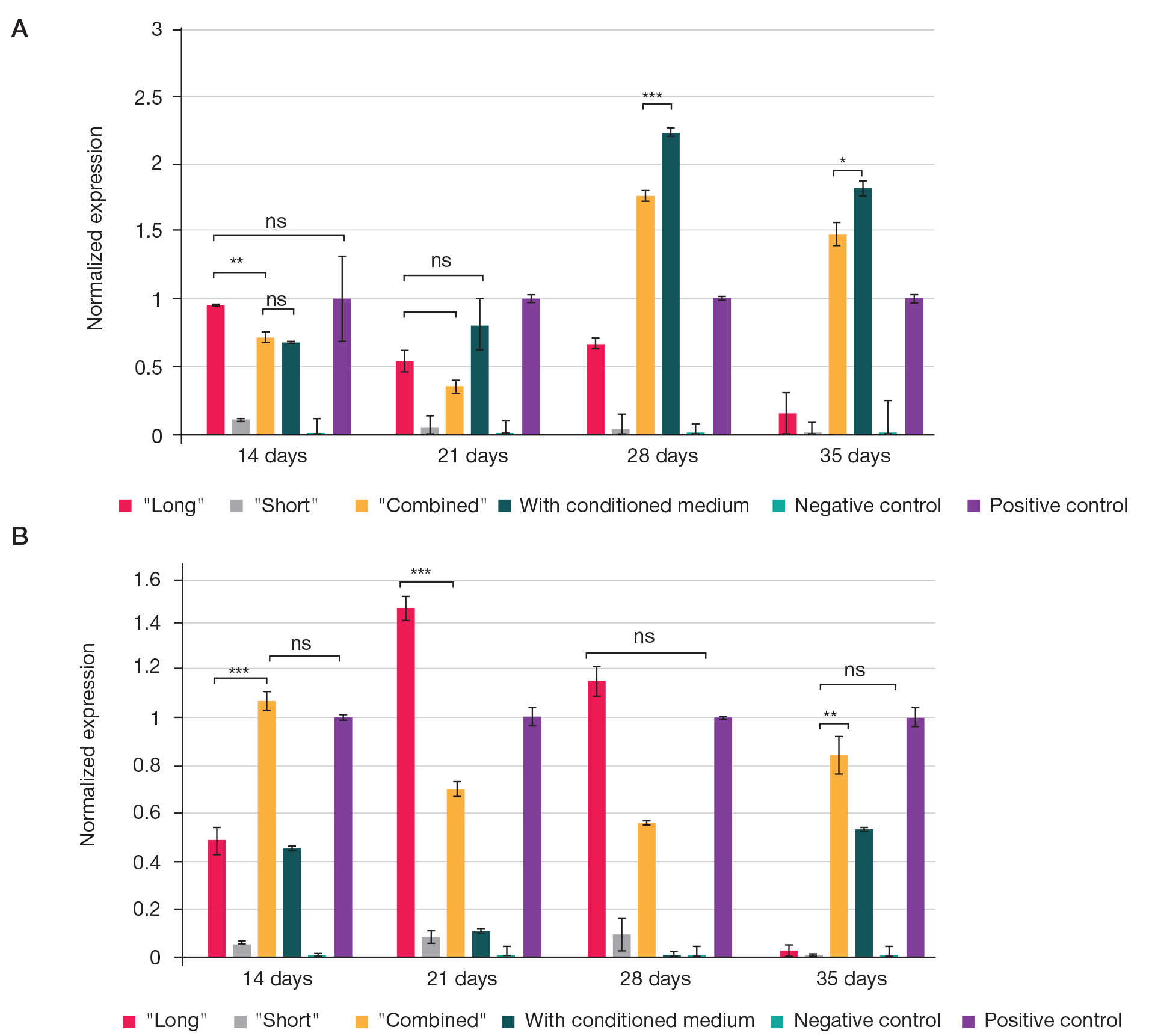
Fig. 5. A. ACAN expression indicators in 3D spheroid cultures of various protocols. B. SOX9 expression indicators. Abscissa, duration of differentiation; ordinate, value of normalized expression. Error bar is the standard deviation. Significance of differences between groups: ns, p > 0.05; * — p < 0.05; * * — p < 0.01; * * * — p < 0.001
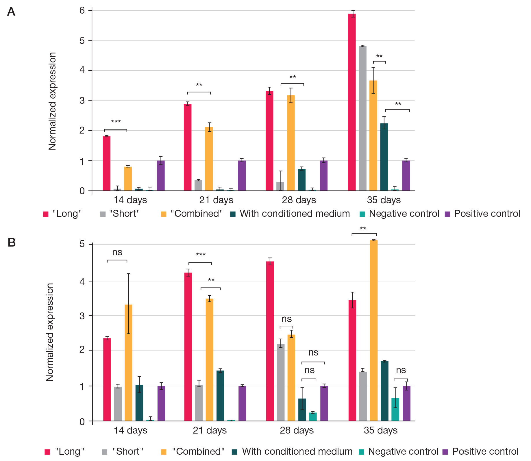
Fig. 6. А. COL1A2 expression indicators in 3D spheroid cultures of various protocols. B. COL2A1 expression indicators. Abscissa, duration of differentiation; ordinate, value of normalized expression. Error bar is the standard deviation. Significance of differences between groups: ns, p > 0.05; * — p < 0.05; * * — p < 0.01; * * * — p < 0.001
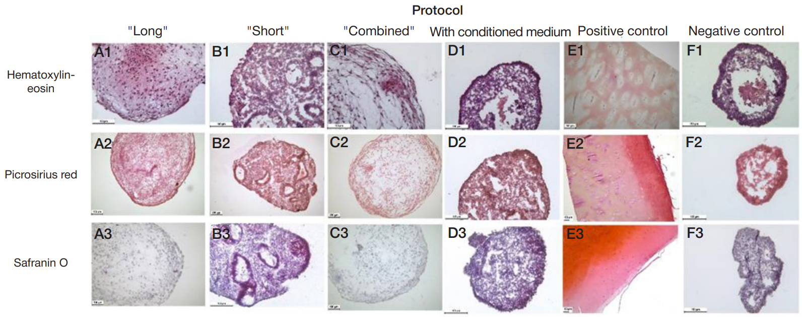
Fig. 7. Histological analysis of 3D spheroid cultures of different differentiation protocols. Differentiation protocols: "long" (A), "short" (B), "combined" (C), with conditioned medium (D). Control groups: positive control (fragments of articular cartilage) (E), negative control (F). Type of histological staining: 1 — hematoxylin-eosin, 2 — picrosirius red, 3 — safranin O
Table 1. Chondrogenic differentiation protocols
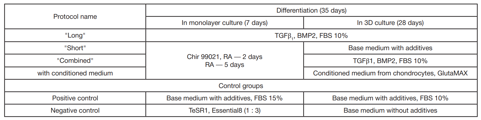
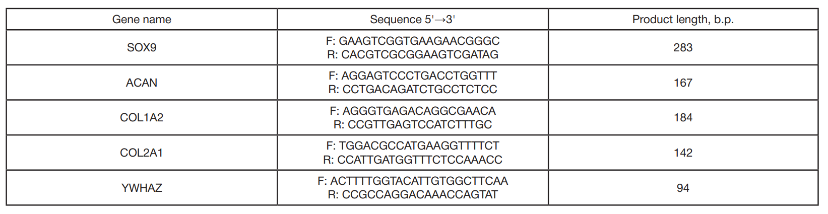
Table 2. Primers used in the work