ORIGINAL RESEARCH

Computational red bone marrow dosimetry phantom of a one-year-old child enabling assessment of exposure due to incorporated beta emitters

About authors

1 Ural Research Center for Radiation Medicine, Chelyabinsk, Russia

2 Chelyabinsk State University, Chelyabinsk, Russia

Correspondence should be addressed: Pavel A. Sharagin
Vorovsky st., 68 A, Chelyabinsk, 454141, Russia; ur.mrcru@nigarahs

About paper

Funding: the work was part of the Federal Target Program "Ensuring Nuclear and Radiation Safety for 2016-2020 and up to 2035", with financial support from the Federal Medical Biological Agency of Russia.

Author contribution: Sharagin PA — data generation, analysis, interpretation, manuscript authoring and editing; Tolstykh EI — study methodology development, manuscript editing; Shishkina EA — conceptualization, manuscript editing.

Received: 2023-06-14 Accepted: 2023-08-23 Published online: 2023-09-26
|
Fig. Segmentation of a hematopoietic site of a one-year-old child's skeleton using the example of humerus. A. Skeleton of a one-year-old child (active hematopoietic sites are highlighted blue). B. Humerus. C. Pattern of division of the bone into BPSs and their dimensions. D. BPS of humerus in voxel representation, a sectional view (black voxels imitate bone, white — BM)
Table 1. AM mass fraction (% of the total AM mass fraction in the skeleton) in the main hematopoietic sites of a one-year-old child's skeleton [15].
Table 2. Chemical composition of the modeled media (all BPS)
Table 3. Spongiosa parameters assumed for BPSs of a one-year-old child [32–42] (CV is given in parentheses, %)
Note: * — spongiosa parameters calculated based on measurements of similar bones or data for other ages; the method of calculation has been described previously [20].
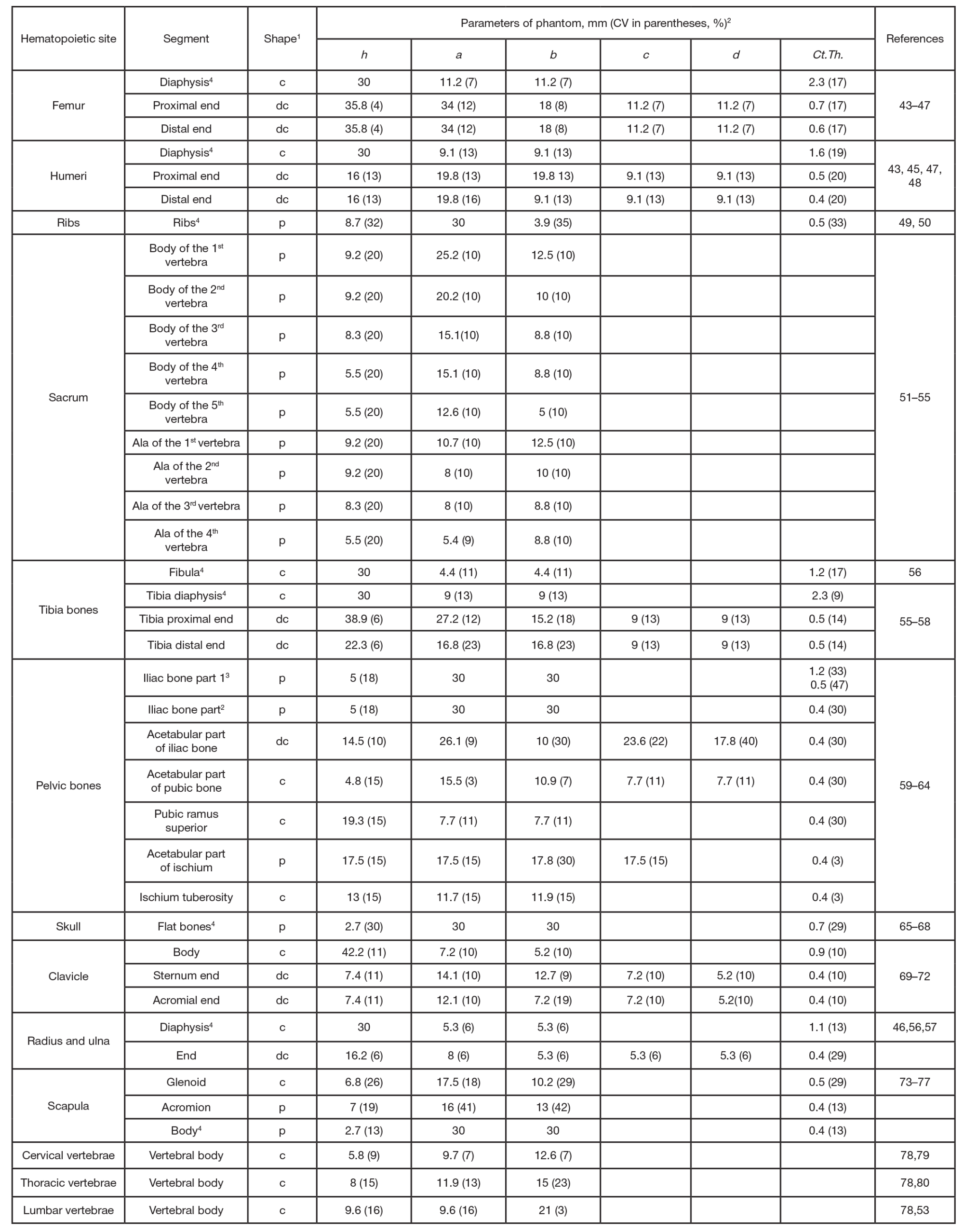
Table 4. Linear dimensions and cortical layer thickness assumed for BPSs of a one-year-old child
Note: 1 — the shape of a phantom was designated as follows: c — cylinder, dc — deformed cylinder, p — rectangular parallelepiped, e — ellipsoid; 2 — dimensions of the BPSs: h — height; a — major axis (c), major axis for a larger base (dc) or side a (r); b — minor axis (c), minor axis for a larger base (dc) or side b (r); c — major axis for smaller base (dc); d — minor axis for smaller base (dc); for ellipsoid (e), a, b, c are the axes; 3 — different cortical layer thickness values were taken for inner (medial) and outer (gluteal) surfaces of this segment of the iliac bone (see Figure); 4 — BPS simulated only part of a modeled bone segment when dimensions thereof significantly exceeded 30 mm, since in such cases from the point of view of dosimetry it makes no sense to model the entire segment [13, 16].
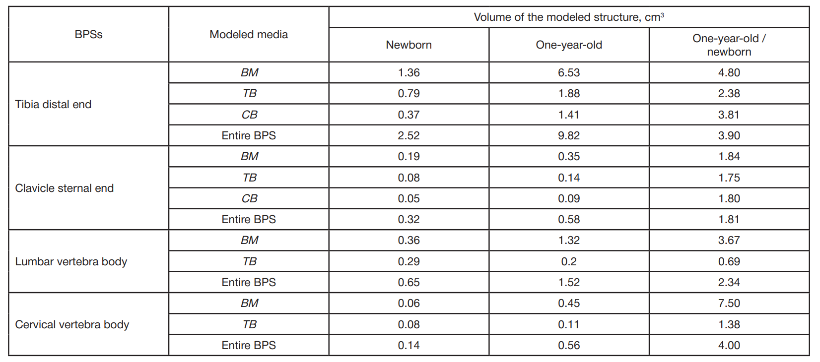
Table 5. Comparison of the volumes of BPSs, newborn and one-year-old child