ORIGINAL RESEARCH

Electron microscopy of the Plasmodium falciparum trophozoites and the tissues these have infected in severe tropical malaria

Solovev AI1, Kapacina VA2, Sokolova MO1, Ariukov AR1, Kovalenko AN1, Uskov AN3, Romanenko VA1
About authors

1 Kirov Military Medical Academy, Saint-Petersburg, Russia

2 Botkin Clinical Infectious Diseases Hospital, Saint-Petersburg, Russia

3 Pediatric Research and Clinical Center of Infectious Diseases of the Federal Medical Biological Agency, Saint Petersburg, Russia

Correspondence should be addressed: Artem R. Ariukov
Akademika Lebedeva, 6, Saint-Petersburg, 194044, Russia; аur.xednay@metra.vokur

About paper

Author contribution: Solovev AI — concept, scientific justification, organization of all types of tests, analysis of the results, manuscript writing; Kapacina VA — data acquisition, practical advising; Sokolova MO, Ariukov AR — sample preparation, light microscopy, analysis of the results, manuscript writing; Kovalenko AN — practical justification, organization of data acquisition, manuscript editing; Uskov AN — concept, scientific advising; Romanenko VA — sample preparation, light microscopy, analysis of the results.

Compliance with ethical standards: the study was approved by the Ethics Committee of the Kirov Military Medical Academy (protocol No. 285 dated 21 November 2023) and conducted in accordance with the principleы of the Declaration of Helsinki (1964) and its subsequent updates.
Received: 2024-05-31 Accepted: 2024-06-26 Published online: 2024-06-29
|
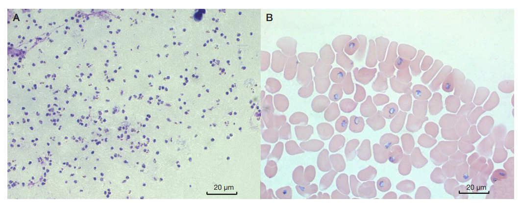
Fig. 1. P. falciparum young (ring) trophozoites in the thick blood smear (А); thin blood smear (B). Romanowsky–Giemsa stain, light microscopy (×1000)
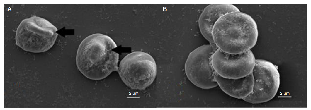
Fig. 2. Scanning electron microscopy of blood specimens. А. Deformation of the erythrocyte cytoplasmic membrane over the trophozoite (arrow). B. Rosetting — formation of conglomerate consisting of infected and uninfected erythrocytes
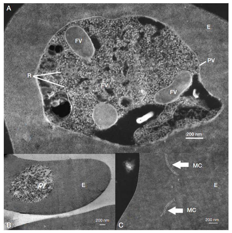
Fig. 3. Transmission electron microscopy: inverted (A, C), direct (B). Random slices of P. falciparum trophozoites located in the erythrocytes. E — erythrocyte; PfP. falciparum; FV — food vacuole; PV — parasitophorous vacuole; R — ribosomes; MC — Maurer's clefts in the infected erythrocyte cytoplasm
Fig. 4. Light microscopy of the autopsy material (hematoxylin and eosin stain, 1000× magnification): cerebral cortex (A); myocardium (B, C). 1 — rosetting in the small capillary lumen; 2 — adhesion of the rosettes of erythrocytes infected with P. falciparum to the capillary wall
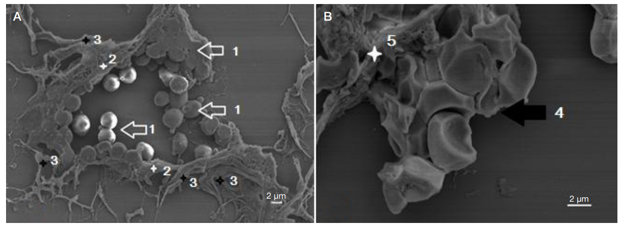
Fig. 5. Scanning electron microscopy of the slices of myocardium (A) and cerebral cortex (B). 1 — adhesion of spherical erythrocytes to the membranes of endothelial cells; 2 — endothelial cells; 3 — fibers of loose fibrous connective tissue along the periphery of the capillary; 4 — resetting in the capillary lumen; 5 — capillary endothelium
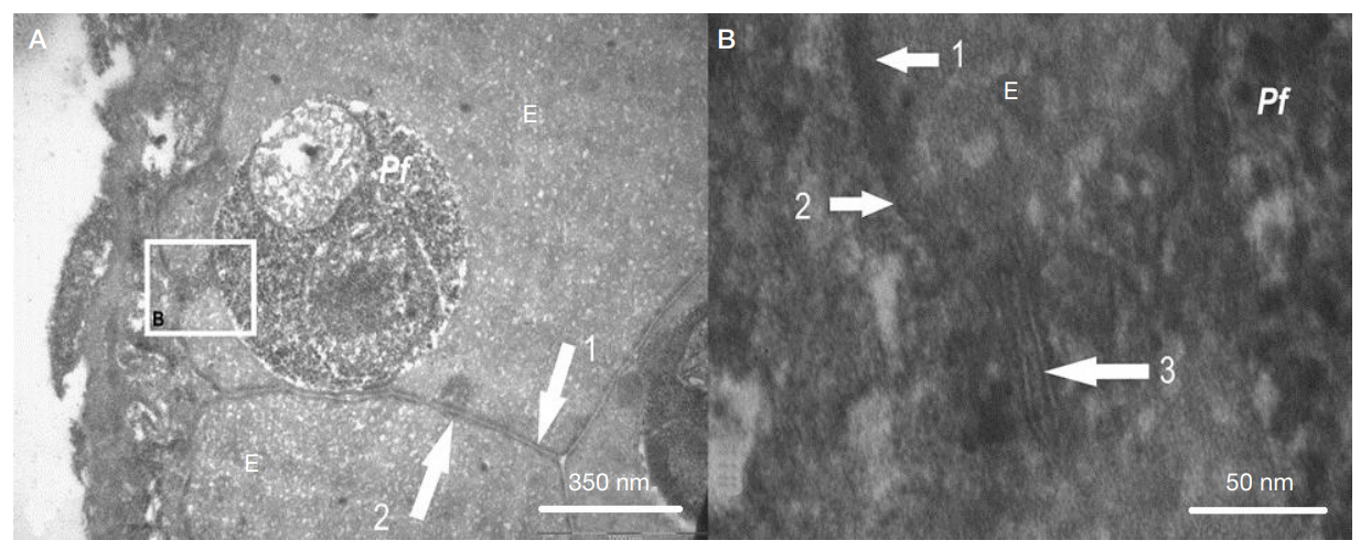
Fig. 6. Transmission electron miscroscopy of brain tissues. A. Slice of mature P. falciparum (Pf) trophozoite inside the erythrocyte (E) fixed on the capillary endothelium surface. B. Abnormal endomembrane system of the infected erythrocyte. 1 — cell membrane of the infected erythrocyte; 2 — cell membrane of the adjacent erythrocyte; 3 — channels of the Maurer's cleft located between the parasitophorous vacuolar membrane and the cell membrane of the affected erythrocyte