METHOD

Identification of phosphonylated peptides using a MALDI target functionalized with lanthanum stearate

Babakov VN1, Gorbunov AYu1, Gladchuk AS2,4, Kalninya YaK2, Shilovskikh VV4, Tomilin NV2, Sukhodolov NG3,4, Radilov AS1, Podolskaya EP2,3
About authors

1 Research Institute of Hygiene, Occupational Pathology and Human Ecology of the Federal Medical Biological Agency, St. Petersburg, Russia

2 Golikov Research Center of Toxicology, St. Petersburg, Russia

3 Institute for Analytical Instrumentation, Russian Academy of Sciences, St. Petersburg, Russia

4 St. Petersburg State University, St. Petersburg, Russia

Correspondence should be addressed: Vladimir Nikolaevich Babakov
Kuzmolovsky, st. Kapitolovo, str. 93, Leningradsraja oblast, Russia; ur.hcepg@vokabab

About paper

Acknowledgments: the authors express their gratitude for technical support to the resource centers Development of Molecular and Cellular Technologies and Geomodel of the Research Park of St. Petersburg State University, and to A.A. Selyutin for the opportunity to use the MALDI mass spectrometer.
Received: 2022-11-11 Accepted: 2023-01-11 Published online: 2023-01-28
|
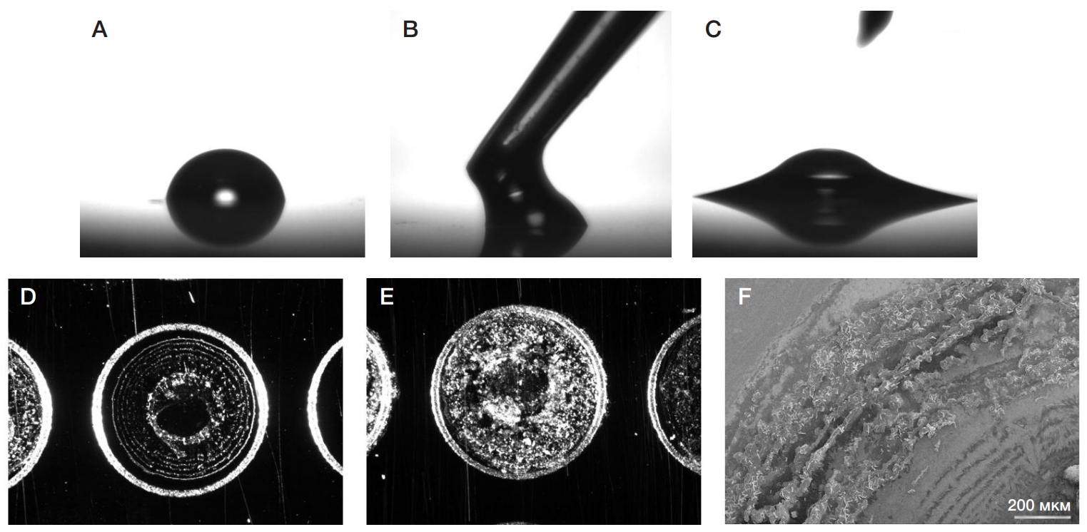
Fig. 1. А. Drop of an aqueous subphase. B. Application of the n-hexane stearic acid solution. C. Distribution of the organic phase over the substrate. D, E. Image of MALDI target spots after deposition of one and six FLa layers (SMZ 1500 stereomicroscope with a DS-2MBWc digital camera (Nikon; Japan)). F. SEM image of six FLa layers formed on the MALDI target (scanning electron microscope S-3400N; Hitachi, Japan)
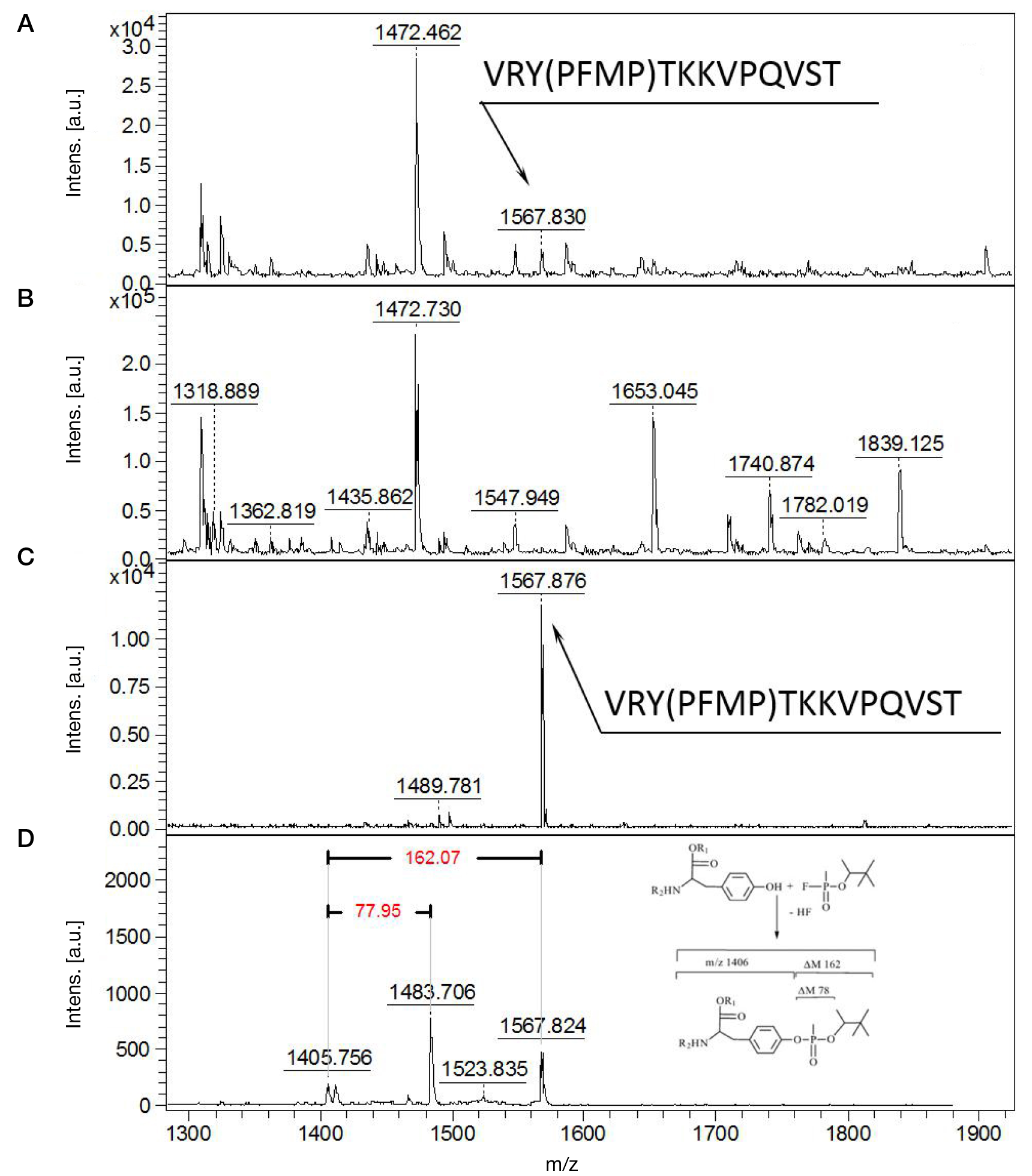
Fig. 2. Mass spectrum of human SA peptic hydrolyzate modified with PFMP at the modified-to-unmodified ratio of 1 : 10. A. Mass spectrum of the original sample. B. Mass spectrum of the unbound fraction. C. Mass spectrum of the adsorbent spot after IMAC. D. PFMP modification identification by neutral loss
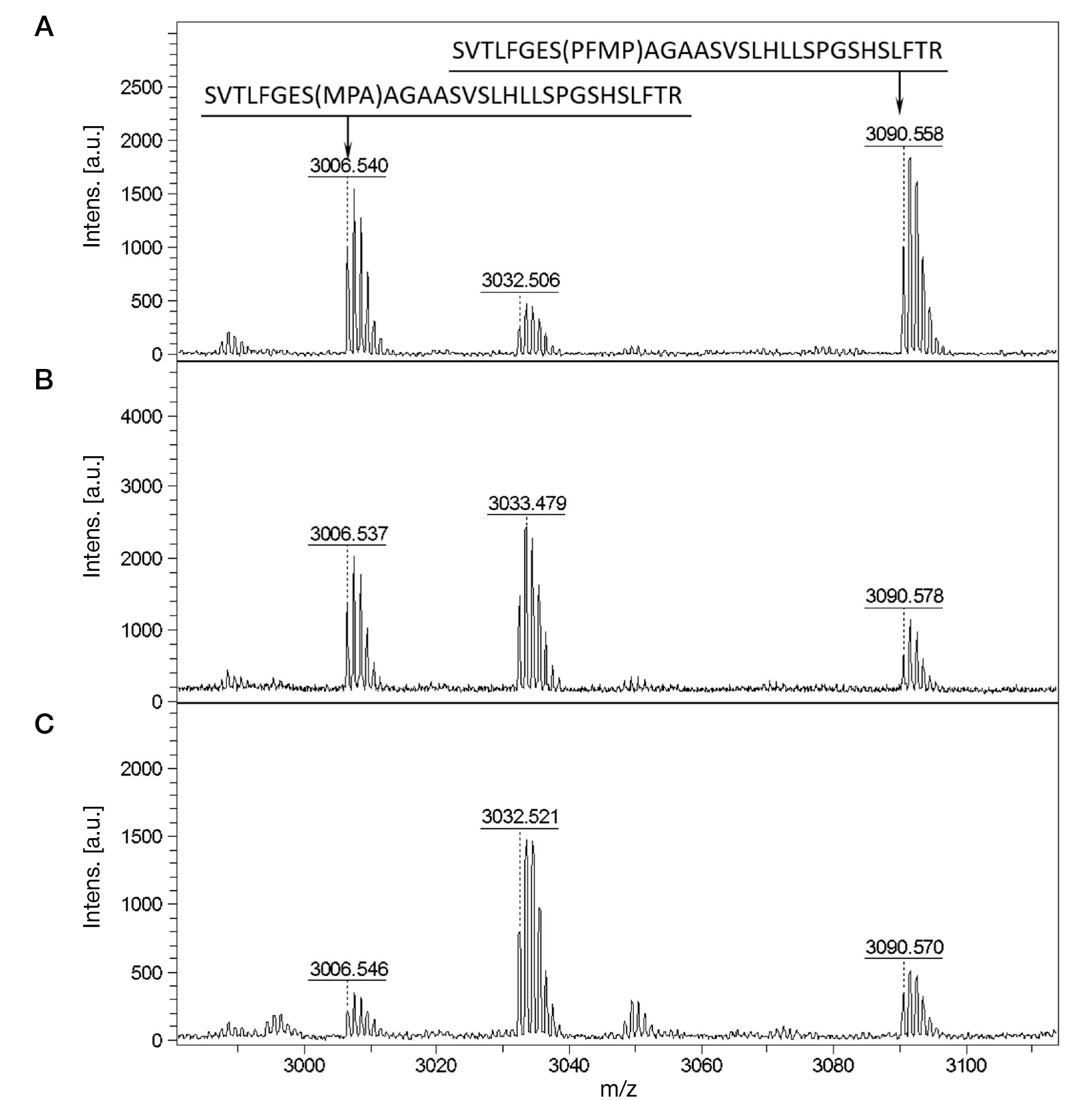
Fig. 3. Results of IMAC(FLa)-MALDI-MS analysis of the tryptic hydrolyzate of BChE (1 mg/ml) modified with PFMP, modified-to-unmodified ratios of 100 : 0 (A); 1 : 10 (B); 1 : 100 (C)
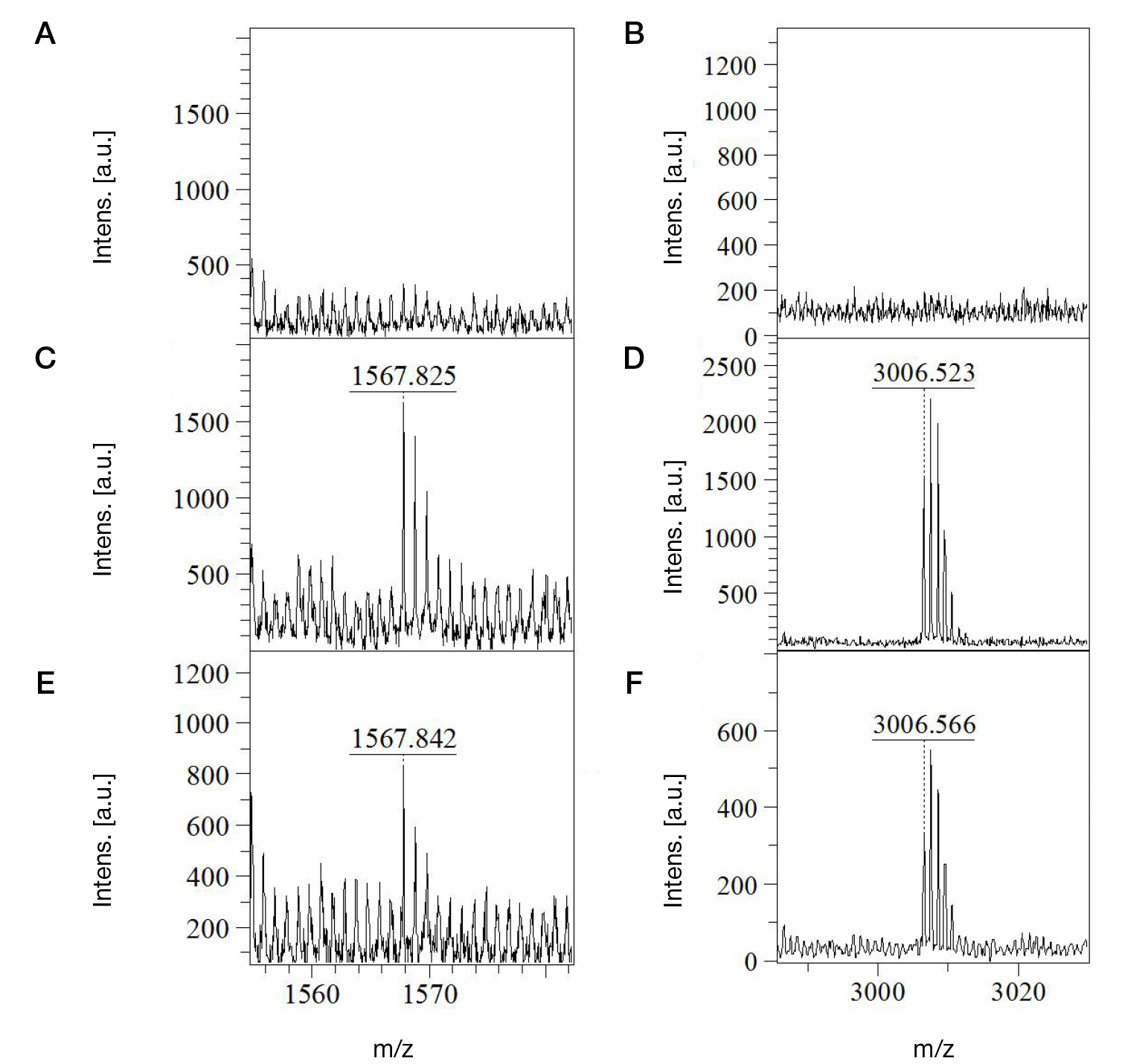
Fig. 4. Results of IMAC(FLa)-MALDI-MS analysis of blood plasma samples incubated with PFMP. The figure shows mass spectra of control samples (A, B); adducts VRY(PFMP)TKKVPQVST and SVTLFGES(MPA)AGAASVSLHLLSPGSHSLFTR, PFMP concentration 10 ng/ml (C, D); VRY(PFMP)TKKVPQVST and SVTLFGES(MPA)AGAASVSLHLLSPGSHSLFTR adducts, PFMP concentration 1 ng/ml (E, F)
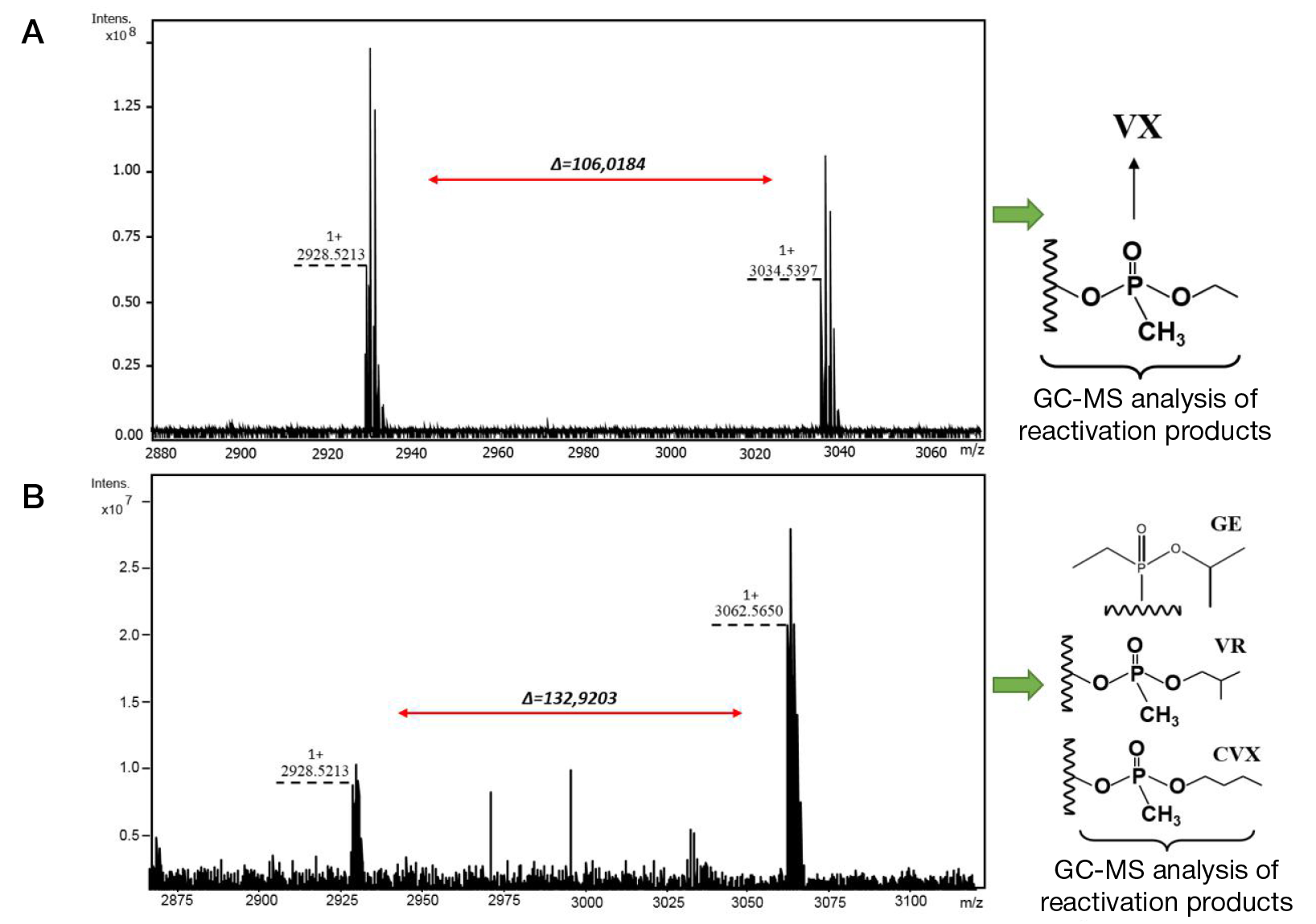
Fig. 5. Results of IMAC(FLa)-MALDI-MS analysis of samples from the Seventh Official OPCW Biomedical Proficiency Test. A. MALDI mass spectrum of tryptic hydrolyzate of BChE modified with VX, signal at m/z 3034.5397, SVTLFGES(Vx)AGAASVSLHLLSPGSHSLFTR. B. MALDI mass spectrum of tryptic hydrolyzate of BChE modified with GE, signal at m/z 3062.5650 - SVTLFGES(GE)AGAASVSLHLLSPGSHSLFTR
Table 1. Results of investigation of selectivity of the developed technique with dilution of PFMP-modified SA with human blood plasma
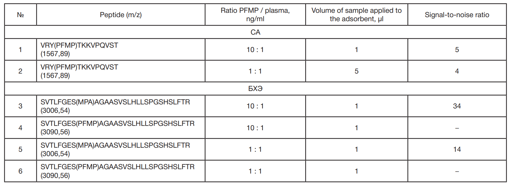
Table 2. Sensitivity of IMAC-MALDI-MS for peptic SA peptides and BChE tryptic peptides modified with PFMP