REVIEW

Role of radiology techniques and hybrid PET-MRI technique in the diagnosis of pharmacoresistant epilepsy

Dolgushin MB, Rostovtseva TM, Duyunova AV, Nadelyaev RV, Beregov MM
About authors

Federal Center of Brain Research and Neurotechnologies of the Federal Medical Biological Agency, Moscow, Russia

Correspondence should be addressed: Tatiana M. Rostovtseva
Ostovityanov, 1, bld. 10, 117513, Moscow, Russia; ur.liam@tavestvotsor

About paper

Funding: the study was performed as part of the research project “Developing Indications for the Use of Hybrid PET-MRI When Planning Surgery in Patients With Epilepsy”, code: 03.02.VY.

Author contribution: all authors contributed significantly to development of the concept, the study and manuscrit writing, they read and approved the final version of the article before publishing. The most significant contributions are distributed as follows: Dolgushin MB — study concept and plan; Rostovtseva TM, Duyunova AV, Nadelyaev RV Duyunova AV, Nadelyaev RV — data acquisition and analysis; Rostovtseva TM, Duyunova AV, Beregov MM — manuscrit writing.
Received: 2023-03-31 Accepted: 2023-05-14 Published online: 2023-06-01
|
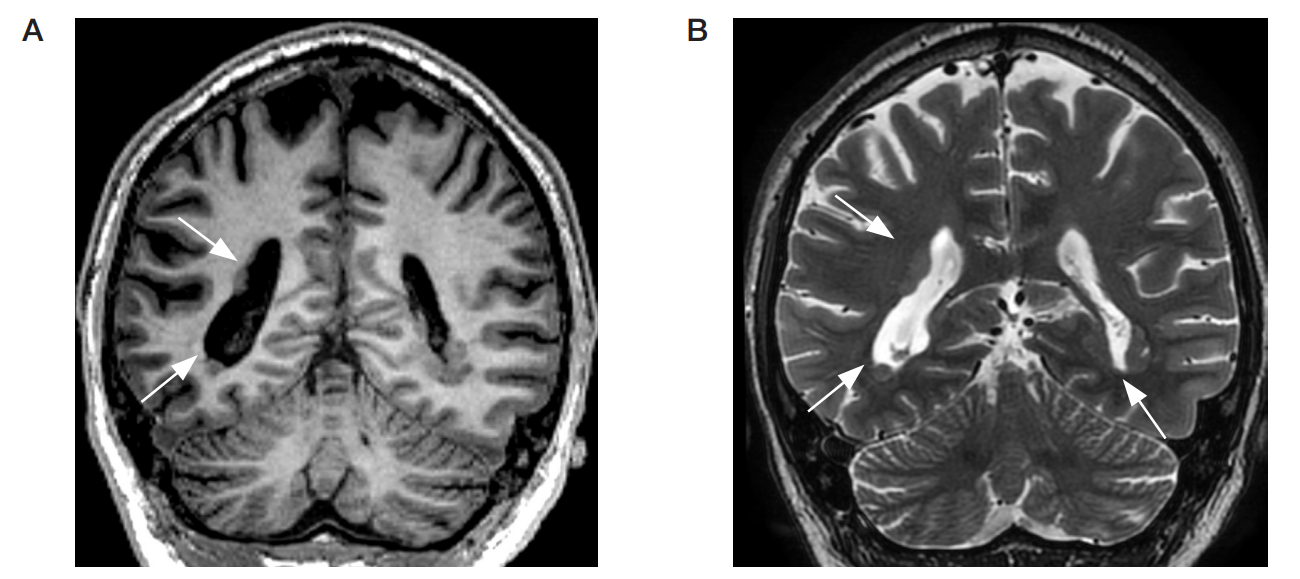
Fig. 1. Brain MRI, oblique coronal scan, T1- (А) and T2-weighted images (B). Multiple areas of subependymal nodular grey matter heterotopia (arrows)
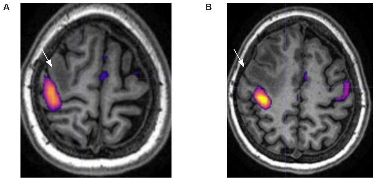
Fig. 2. Functional MRI. Motor cortex mapping in patient with refractory epilepsy (А, B). Distribution of the areas showing activation when moving the left fingers is marked by the arrowhead, the epileptogenic zone is marked by the arrow
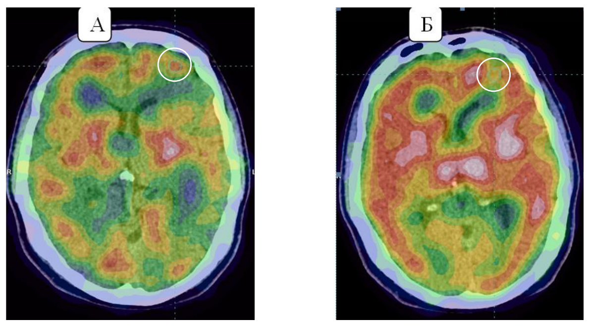
Fig. 3. SPECT perfusion maps (RPA 99mTc-Teoxim) of the patient with refractory epilepsy performed in ictal (A) and interictal (B) phases. The annular area in the left middle frontal gyrus markes the zone of hyperperfusion in the images obtained during the ictal phase that corresponds to the hypoperfusion zone in the images obtained during the interictal phase
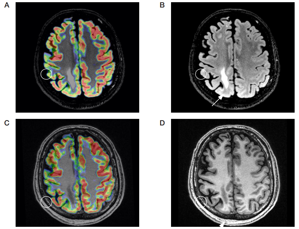
Fig. 4. 18F-FDG-PET-MRI of the brain in patient with pharmacoresistant epilepsy and unsatisfactory outcome of epilepsy surgical treatment. А, C: area of hypometabolism in the dorsolateral cortex of the right frontal lobe (circle). Brain MRI: axial FLAIR (B), IR-FSPGR (D). Focal cortical thickening with FLAIR hyperintensity in the area of hypometabolism (circle). Postsurgical structural brain changes in the paramedian area of the right parietal lobe (arrow)